
Page 806 - Textbook of Pathology, 6th Edition
P. 806
790 stage, mycosis fungoides may disseminate to the lymph
nodes and other organs. Clinically, mycosis fungoides may
manifest in 3 stages:
i) Premycotic stage in which the lesions are erythematous,
red-brown, scaly and pruritic, resembling eczema or
psoriasis.
ii) Infiltrative stage has slightly elevated, bluish-red, firm
plaques.
iii) Fungoid (Tumour) stage is characterised by red-brown
nodules of tumour which often undergo ulceration.
The etiology of mycosis fungoides or CTCL has been
found to be the same as for adult T cell lymphoma-leukaemia
syndrome which is human T cell-leukaemia virus-I (HTLV-
I) as discussed on page 227.
Sézary syndrome is a variant of CTCL, often due to
dissemination of underlying CTCL to the blood and infil-
tration into the skin causing generalised erythroderma,
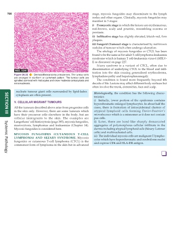
Figure 26.32 Dermatofibrosarcoma protuberans. The tumour cells lymphadenopathy and hepatosplenomegaly.
are arranged in storiform or cartwheel pattern. The tumour cells are
spindled admixed with histiocytes and show moderate anisocytosis and The condition is found more frequently beyond 4th
anisonucleosis. decade of life. Lesions may affect different body surfaces but
often involve the trunk, extremities, face and scalp.
nucleate tumour giant cells surrounded by lipid-laden Histologically, the condition has the following charac-
cytoplasm are often present.
teristics:
i) Initially, lower portion of the epidermis contains
V. CELLULAR MIGRANT TUMOURS
hyperchromatic enlarged lymphocytes. In about half the
All the tumours described above arise from progenitor cells cases, there is formation of intraepidermal clusters of
in the skin only. However, there are some tumours which atypical lymphoid cells forming Darier-Pautrier’s
have their precursor cells elsewhere in the body, but are microabscesses which is a misnomer as it does not contain
SECTION III
cellular immigrants to the skin. The examples are pus cells.
Langerhans’ cell histiocytosis (page 385), mycosis fungoides, ii) Later, there are band-like sharply demarcated
mastocytosis, lymphomas and leukaemias (Chapter 14). aggregates of polymorphous cellular infiltrate in the
Mycosis fungoides is considered here. dermis including atypical lymphoid cells (Sézary-Lutzner
cells) and multinucleated cells.
MYCOSIS FUNGOIDES (CUTANEOUS T-CELL iii) The individual mycosis cells are malignant T lympho-
LYMPHOMA) AND SEZARY SYNDROME. Mycosis cytes which have hyperchromatic and cerebriform nuclei
fungoides or cutaneous T-cell lymphoma (CTCL) is the and express CD4 and HLA-DR antigen.
commonest form of lymphoma in the skin but in advanced
❑
Systemic Pathology