
Page 817 - Textbook of Pathology, 6th Edition
P. 817
801
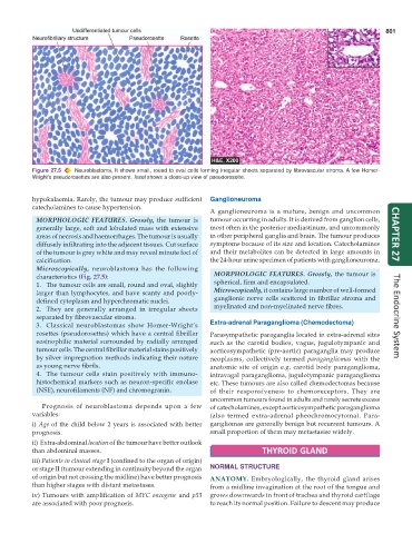
Figure 27.5 Neuroblastoma, It shows small, round to oval cells forming irregular sheets separated by fibrovascular stroma. A few Homer-
Wright’s pseudorosettes are also present. Inset shows a close-up view of pseudorosette.
hypokalaemia. Rarely, the tumour may produce sufficient Ganglioneuroma
catecholamines to cause hypertension.
A ganglioneuroma is a mature, benign and uncommon
MORPHOLOGIC FEATURES. Grossly, the tumour is tumour occurring in adults. It is derived from ganglion cells,
generally large, soft and lobulated mass with extensive most often in the posterior mediastinum, and uncommonly
areas of necrosis and haemorrhages. The tumour is usually in other peripheral ganglia and brain. The tumour produces CHAPTER 27
diffusely infiltrating into the adjacent tissues. Cut surface symptoms because of its size and location. Catecholamines
of the tumour is grey white and may reveal minute foci of and their metabolites can be detected in large amounts in
calcification. the 24-hour urine specimen of patients with ganglioneuroma.
Microscopically, neuroblastoma has the following
characteristics (Fig. 27.5): MORPHOLOGIC FEATURES. Grossly, the tumour is
1. The tumour cells are small, round and oval, slightly spherical, firm and encapsulated.
larger than lymphocytes, and have scanty and poorly- Microscopically, it contains large number of well-formed
defined cytoplasm and hyperchromatic nuclei. ganglionic nerve cells scattered in fibrillar stroma and
2. They are generally arranged in irregular sheets myelinated and non-myelinated nerve fibres.
separated by fibrovascular stroma. The Endocrine System
3. Classical neuroblastomas show Homer-Wright’s Extra-adrenal Paraganglioma (Chemodectoma)
rosettes (pseudorosettes) which have a central fibrillar Parasympathetic paraganglia located in extra-adrenal sites
eosinophilic material surrounded by radially arranged such as the carotid bodies, vagus, jugulotympanic and
tumour cells. The central fibrillar material stains positively aorticosympathetic (pre-aortic) paraganglia may produce
by silver impregnation methods indicating their nature neoplasms, collectively termed paragangliomas with the
as young nerve fibrils. anatomic site of origin e.g. carotid body paraganglioma,
4. The tumour cells stain positively with immuno- intravagal paraganglioma, jugulotympanic paraganglioma
histochemical markers such as neuron-specific enolase etc. These tumours are also called chemodectomas because
(NSE), neurofilaments (NF) and chromogranin. of their responsiveness to chemoreceptors. They are
uncommon tumours found in adults and rarely secrete excess
Prognosis of neuroblastoma depends upon a few of catecholamines, except aorticosympathetic paraganglioma
variables: (also termed extra-adrenal pheochromocytoma). Para-
i) Age of the child below 2 years is associated with better gangliomas are generally benign but recurrent tumours. A
prognosis. small proportion of them may metastasise widely.
ii) Extra-abdominal location of the tumour have better outlook
than abdominal masses. THYROID GLAND
iii) Patients in clinical stage I (confined to the organ of origin)
or stage II (tumour extending in continuity beyond the organ NORMAL STRUCTURE
of origin but not crossing the midline) have better prognosis ANATOMY. Embryologically, the thyroid gland arises
than higher stages with distant metastases. from a midline invagination at the root of the tongue and
iv) Tumours with amplification of MYC oncogene and p53 grows downwards in front of trachea and thyroid cartilage
are associated with poor prognosis. to reach its normal position. Failure to descent may produce