
Page 848 - Textbook of Pathology, 6th Edition
P. 848
832
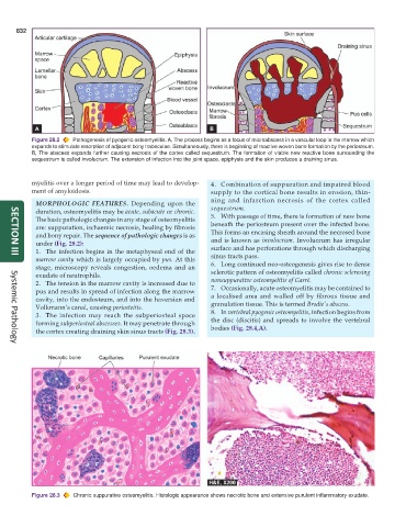
Figure 28.2 Pathogenesis of pyogenic osteomyelitis. A, The process begins as a focus of microabscess in a vascular loop in the marrow which
expands to stimulate resorption of adjacent bony trabeculae. Simultaneously, there is beginning of reactive woven bone formation by the periosteum.
B, The abscess expands further causing necrosis of the cortex called sequestrum. The formation of viable new reactive bone surrounding the
sequestrum is called involucrum. The extension of infection into the joint space, epiphysis and the skin produces a draining sinus.
myelitis over a longer period of time may lead to develop- 4. Combination of suppuration and impaired blood
ment of amyloidosis. supply to the cortical bone results in erosion, thin-
ning and infarction necrosis of the cortex called
MORPHOLOGIC FEATURES. Depending upon the
sequestrum.
duration, osteomyelitis may be acute, subacute or chronic. 5. With passage of time, there is formation of new bone
The basic pathologic changes in any stage of osteomyelitis
are: suppuration, ischaemic necrosis, healing by fibrosis beneath the periosteum present over the infected bone.
and bony repair. The sequence of pathologic changes is as This forms an encasing sheath around the necrosed bone
under (Fig. 28.2): and is known as involucrum. Involucrum has irregular
1. The infection begins in the metaphyseal end of the surface and has perforations through which discharging
marrow cavity which is largely occupied by pus. At this sinus tracts pass.
SECTION III
stage, microscopy reveals congestion, oedema and an 6. Long continued neo-osteogenesis gives rise to dense
exudate of neutrophils. sclerotic pattern of osteomyelitis called chronic sclerosing
2. The tension in the marrow cavity is increased due to nonsuppurative osteomyelitis of Garré.
pus and results in spread of infection along the marrow 7. Occasionally, acute osteomyelitis may be contained to
cavity, into the endosteum, and into the haversian and a localised area and walled off by fibrous tissue and
granulation tissue. This is termed Brodie’s abscess.
Volkmann’s canal, causing periosteitis. 8. In vertebral pyogenic osteomyelitis, infection begins from
3. The infection may reach the subperiosteal space
forming subperiosteal abscesses. It may penetrate through the disc (discitis) and spreads to involve the vertebral
the cortex creating draining skin sinus tracts (Fig. 28.3). bodies (Fig. 28.4,A).
Systemic Pathology
Figure 28.3 Chronic suppurative osteomyelitis. Histologic appearance shows necrotic bone and extensive purulent inflammatory exudate.