
Page 846 - Textbook of Pathology, 6th Edition
P. 846
830
Chapter 28 The Musculoskeletal System
Chapter 28
SKELETAL SYSTEM line phosphatase (other being hepatic alkaline phosphatase) is
a marker for osteoblastic activity. Its levels are raised in
The skeleton consists of cartilage and bone. Cartilage has a puberty during period of active bone growth and in
role in growth and repair of bone, and in the adults forms pathologic conditions associated with high osteoblastic
the articular skeleton responsible for movement of joints. activity such as in fracture repair and Paget’s disease of the
Bone is a specialised form of connective tissue which bone.
performs the function of providing mechanical support and 2. Osteocytes. Osteocytes are those osteoblasts which get
is also a mineral reservoir for calcium homeostasis. There incorporated into the bone matrix during its synthesis. Osteo-
are 206 bones in the human body, and depending upon their cytes are found within small spaces called lacunae lying in
size and shape may be long, flat, tubular etc. the bone matrix. The distribution of the osteocytic lacunae is
a reliable parameter for distinguishing between woven and
NORMAL STRUCTURE OF BONE lamellar bone.
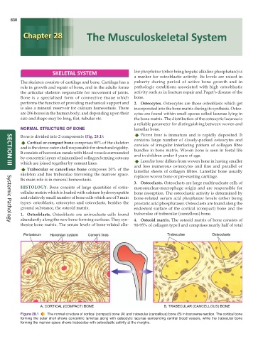
Bone is divided into 2 components (Fig. 28.1): Woven bone is immature and is rapidly deposited. It
Cortical or compact bone comprises 80% of the skeleton contains large number of closely-packed osteocytes and
and is the dense outer shell responsible for structural rigidity. consists of irregular interlacing pattern of collagen fibre
bundles in bone matrix. Woven bone is seen in foetal life
It consists of haversian canals with blood vessels surrounded and in children under 4 years of age.
by concentric layers of mineralised collagen forming osteons
Lamellar bone differs from woven bone in having smaller
which are joined together by cement lines. and less numerous osteocytes and fine and parallel or
SECTION III
Trabecular or cancellous bone composes 20% of the lamellar sheets of collagen fibres. Lamellar bone usually
skeleton and has trabeculae traversing the marrow space. replaces woven bone or pre-existing cartilage.
Its main role is in mineral homeostasis.
3. Osteoclasts. Osteoclasts are large multinucleate cells of
HISTOLOGY. Bone consists of large quantities of extra- mononuclear-macrophage origin and are responsible for
cellular matrix which is loaded with calcium hydroxyapatite bone resorption. The osteoclastic activity is determined by
and relatively small number of bone cells which are of 3 main bone-related serum acid phosphatase levels (other being
types: osteoblasts, osteocytes and osteoclasts, besides the prostatic acid phosphatase). Osteoclasts are found along the
ground substance, the osteoid matrix. endosteal surface of the cortical (compact) bone and the
1. Osteoblasts. Osteoblasts are uninucleate cells found trabeculae of trabecular (cancellous) bone.
abundantly along the new bone-forming surfaces. They syn- 4. Osteoid matrix. The osteoid matrix of bone consists of
Systemic Pathology
thesise bone matrix. The serum levels of bone-related alka- 90-95% of collagen type I and comprises nearly half of total
Figure 28.1 The normal structure of cortical (compact) bone (A) and trabecular (cancellous) bone (B) in transverse section. The cortical bone
forming the outer shell shows concentric lamellae along with osteocytic lacunae surrounding central blood vessels, while the trabecular bone
forming the marrow space shows trabeculae with osteoclastic activity at the margins.