
Page 852 - Textbook of Pathology, 6th Edition
P. 852
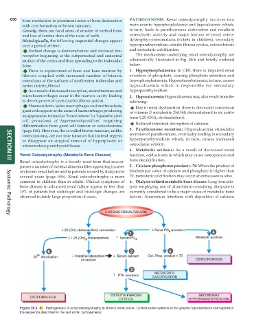
836   bone rarefaction to prominent areas of bone destruction  PATHOGENESIS.  Renal osteodystrophy involves two
            with cyst formation or brown tumours.              main events: hyperphosphataemia and hypocalcaemia which,
            Grossly, there are focal areas of erosion of cortical bone  in turn, leads to parathormone elaboration and resultant
            and loss of lamina dura at the roots of teeth.     osteoclastic activity and major lesions of renal osteo-
            Histologically, the following sequential changes appear  dystrophy—osteomalacia (rickets in children), secondary
            over a period of time:                             hyperparathyroidism, osteitis fibrosa cystica, osteosclerosis
               Earliest change is demineralisation and increased bone  and metastatic calcification.
            resorption beginning at the subperiosteal and endosteal  The mechanisms underlying renal osteodystrophy are
            surface of the cortex and then spreading to the trabecular  schematically illustrated in Fig. 28.6 and briefly outlined
            bone.                                              below:
               There is replacement of bone and bone marrow by  1. Hyperphosphataemia: In CRF, there is impaired renal
            fibrosis coupled with increased number of bizarre  excretion of phosphate, causing phosphate retention and
            osteoclasts at the surfaces of moth-eaten trabeculae and  hyperphosphataemia. Hyperphosphataemia, in turn, causes
            cortex (osteitis fibrosa).                         hypocalcaemia which is responsible for secondary
               As a result of increased resorption, microfractures and  hyperparathyroidism.
            microhaemorrhages occur in the marrow cavity leading  2. Hypocalcaemia: Hypocalcaemia may also result from the
            to development of cysts (osteitis fibrosa cystica).  following:
               Haemosiderin-laden macrophages and multinucleate   Due to renal dysfunction, there is decreased conversion
            giant cells appear at the areas of haemorrhages producing  of vitamin D metabolite 25(OH) cholecalciferol to its active
            an appearance termed as ‘brown tumour’ or ‘reparative giant  form 1,25 (OH) cholecalciferol.
                                                                            2
            cell granuloma of hyperparathyroidism’ requiring      Reduced intestinal absorption of calcium.
            differentiation from giant cell tumour or osteoclastoma
            (page 846). However, the so-called brown tumours, unlike  3. Parathormone secretion: Hypocalcaemia stimulates
            osteoclastoma, are not true tumours but instead regress  secretion of parathormone, eventually leading to secondary
            or disappear on surgical removal of hyperplastic or  hyperparathyroidism which, in turn, causes increased
            adenomatous parathyroid tissue.                    osteoclastic activity.
                                                               4. Metabolic acidosis: As a result of decreased renal
           Renal Osteodystrophy (Metabolic Bone Disease)       function, acidosis sets in which may cause osteoporosis and
           Renal osteodystrophy is a loosely used term that encom-  bone decalcification.
     SECTION III
           passes a number of skeletal abnormalities appearing in cases  5. Calcium phosphorus product > 70: When the product of
           of chronic renal failure and in patients treated by dialysis for  biochemical value of calcium and phosphate is higher than
           several years (page 656). Renal osteodystrophy is more  70, metastatic calcification may occur at extraosseous sites.
           common in children than in adults. Clinical symptoms of  6. Dialysis-related metabolic bone disease: Long-term dia-
           bone disease in advanced renal failure appear in less than  lysis employing use of aluminium-containing dialysate is
           10% of patients but radiologic and histologic changes are  currently considered to be a major cause of metabolic bone
           observed in fairly large proportion of cases.       lesions. Aluminium interferes with deposition of calcium


     Systemic Pathology


           Figure 28.6  Pathogenesis of renal osteodystrophy in chronic renal failure. Circled serial numbers in the graphic representation correspond to
           the sequence described in the text under pathogenesis.