


Page 101 - Textbook of Pathology, 6th Edition
P. 101
immunoglobulin, or λ light chain, or κ light chain, or rarely Based on cause, into primary (with unknown cause and 85
heavy chains. This takes place by monoclonal proliferation the deposition is in the disease itself) and secondary (as a
of plasma cells, B lymphocytes, or their precursors. complication of some underlying known disease)
3. Partial degradation in the form of limited proteolysis of amyloidosis.
larger protein molecules occurs in macrophages that are Based on extent of amyloid deposition, into systemic
anatomically closely associated with AL amyloid. (generalised) involving multiple organs and localised CHAPTER 4
4. Non-fibrillar components like AP and GAGs play some role amyloidosis involving one or two organs or sites.
in folding and aggregation of fibril proteins. Based on histological basis, into pericollagenous (corres-
ponding in distribution to primary amyloidosis), and
Deposition of AA Amyloid
perireticulin (corresponding in distribution to secondary
1. AA amyloid is directly related to SAA levels, a high- amyloidosis).
density lipoprotein. SAA is synthesised by the liver in Based on clinical location, into pattern I (involving
response to cytokines, notably interleukin 1 and 6, released tongue, heart, bowel, skeletal and smooth muscle, skin and
from activated macrophages. nerves), pattern II (principally involving liver, spleen, kidney
2. The levels of SAA are elevated in long-standing tissue and adrenals) and mixed pattern (involving sites of both
destruction e.g. in chronic inflammation, cancers. However, pattern I and II).
SAA levels in isolation do not always lead to AA amyloid. Based on tissues in which amyloid is deposited, into
3. As in AL amyloid, partial degradation in the form of limited mesenchymal (organs derived from mesoderm) and
proteolysis takes place in reticuloendothelial cells. parenchymal (organs derived from ectoderm and endoderm)
4. In AA amyloid, a significant role is played by another
glycoprotein, amyloid enhancing factor (AEF). The exact amyloidosis. Immunopathology Including Amyloidosis
composition of AEF is not known. AEF is elaborated in Based on precursor biochemical proteins, into specific
chronic inflammation, cancer and familial Mediterranean type of serum amyloid proteins.
fever. On the basis of experimental induction of AA amyloid, With availability of biochemical composition of various
AEF has been shown to accelerate AA amyloid deposition. forms of amyloid and diverse clinical settings in which these
Possibly, AEF acts as a nidus for deposition of fibrils in AA specific biochemical forms of amyloid are deposited, a
amyloid. clinicopathologic classification has been proposed which is
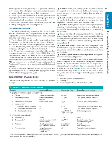
5. As in AL amyloid, there is a role of AP component and widely acceptable (Table 4.10). According to this
glycosaminoglycans in the fibril protein aggregation and to classification, amyloidosis can be divided into 2 major
protect it from disaggregation again. categories and their subtypes depending upon clinical
settings:
CLASSIFICATION OF AMYLOIDOSIS
A. Systemic (generalised) amyloidosis:
Over the years, amyloidosis has been classified in a number 1. Primary (AL)
of ways: 2. Secondary/reactive/ inflammatory (AA)
TABLE 4.10: Classification of Amyloidosis.
Category Associated Disease Biochemical Type Organs Commonly Involved
A. SYSTEMIC (GENERALISED) AMYLOIDOSIS
1. Primary Plasma cell dyscrasias AL type Heart, bowel, skin, nerves, kidney
2. Secondary (Reactive) Chronic inflammation, AA type Liver, spleen, kidneys, adrenals
cancers
3. Haemodialysis-associated Chronic renal failure Aβ 2 M Synovium, joints, tendon sheaths
4. Heredofamilial
i. Hereditary polyneuropathies — ATTR Peripheral and autonomic nerves, heart
ii. Familial Mediterranean fever — AA type Liver, spleen, kidneys, adrenals
iii. Rare hereditary forms — AApoAI, AGel Systemic amyloidosis
ALys, AFib, ACys
B. LOCALISED AMYLOIDOSIS
1. Senile cardiac Senility ATTR Heart
2. Senile cerebral Alzheimer’s, transmissible Aβ, APrP Cerebral vessels, plaques,
encephalopathy neurofibrillary tangles
3. Endocrine Medullary carcinoma Procalcitonin Thyroid
type 2 diabetes mellitus Proinsulin Islets of Langerhans
4. Tumour-forming Lungs, larynx, skin, AL Respective anatomic location
urinary bladder,
tongue, eye
(AL= Amyloid light chain; AA= Amyloid-associated protein; Aβ 2 M= Amyloid β 2 -microglobulin; ATTR= Amyloid transthyretin; APrP=Amyloid of
prion proteins, Aβ= β-amyloid protein).

