
Page 48 - TI Journal 18-1
P. 48
42 TATE
research has focused on the potential of rTMS to
alleviate symptoms associated with a wide range of
neurological and psychiatric conditions, including
schizophrenic hallucinations, tinnitus, anxiety, neu-
rodegenerative disorders, and chronic pain (89)
The use of MRI in combination with TMS has been
the subject of significant interest (87). TMS can be
delivered before, during, or after MRI. This flexibil-
ity can be used to establish causality or correlations
between imaging changes and behavior previously
noted on MRI. Frameless stereotactic systems use
structural MRI data to precisely deliver TMS in a
specific location (75). While these multimodal studies
increase specificity regarding focal brain targets for
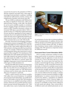
treatment, they do not explain the full effect of TMS Figure 2. Participant working on training task while wearing
within the brain and are significantly limited by the tDCS electrodes.
interconnected nature of brain networks and the
diffuse activation that occurs with TMS (61,89). its mechanisms of action due to technical problems
Recently, functional neuroimaging techniques with coil miniaturization. In addition, the fact that
using positron emission tomography (PET) and func- TMS has already received FDA approval reduces the
tional MRI (fMRI) have been employed to study the incentive for further animal work (89). To address
effects of TMS. These studies support the efficacy of these limitations, human studies combining neuro-
TMS methods in eliciting noticeable functional brain imaging with TMS represent an important avenue
changes. For example, rTMS has been linked to sub- for additional research.
cortical dopamine release with connections to cortical
projection fibers using PET (79). Simultaneous TMS- Transcranial Direct Current Stimulation (tDCS)
fMRI studies also yield valuable information that has tDCS is another noninvasive method of modu-
both high temporal and spatial resolution. These lating neural activity via increases or decreases in
data demonstrate the ability of the brain to adapt excitability using the application of weak electrical
to inhibitory TMS effects in a specific region and currents (0.5–2 mA) to the brain with two or more
highlight compensatory neural connections outside electrodes. The current enters the head from the
of the region of TMS stimulation (4). anode(s), travels through the tissue, and flows back
Resting state fMRI shows promise in demonstrat- to the cathode(s). As the current flows between the
ing functional connectivity changes induced by TMS electrodes, it is believed to modulate neural activity
(86). This may allow the use of resting state connec- beneath the electrodes, and the effects are dependent
tivity as a surrogate marker for TMS effectiveness on the direction, strength, and duration of the current
in diseases such as chronic pain, where the clinical (48). At moderate levels of current intensity (e.g., 1
effect size of treatment is small (58). mA), neurons influenced by the anodal (+) stimula-
While a useful research and clinical modality, tion appear to increase neuronal excitability via slight
TMS suffers from several limitations. In modeling depolarization. In contrast, neurons that are influ-
structural-functional relationships, one notable draw- enced by the cathode (-) stimulation are inhibited
back in TMS studies is the fact that TMS stimulates by hyperpolarization (52,67). However, higher cur-
neural tissue in an amplified and perhaps artificial rent strengths (e.g., 2 mA) have been shown to cause
manner, which may not accurately represent conven- increases in excitability in brain tissues influenced
tional neuronal network firing patterns (22). TMS by both anode and cathode. Thus, cortical effects
also suffers from a lack of animal data describing of different anode and cathode placements are not