Page 226 - Inventions - A Visual Encyclopedia (DK - Smithsonian)
P. 226

Looking inside


          Before 1895, looking inside a patient’s body meant cutting
          it open. The discovery of X-rays by the German scientist

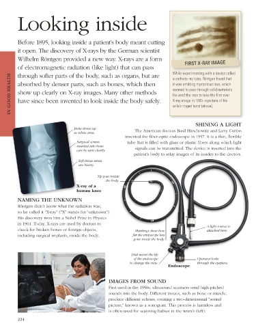
          Wilhelm Röntgen provided a new way. X-rays are a form                           FIRST X-RAY IMAGE
          of electromagnetic radiation (like light) that can pass                   While experimenting with a device called

          through softer parts of the body, such as organs, but are
      IN GOOD HEALTH  absorbed by denser parts, such as bones, which then           it was emitting mysterious rays, which
                                                                                    a cathode ray tube, Röntgen found that
                                                                                    seemed to pass through solid materials.
          show up clearly on X-ray images. Many other methods
                                                                                    He used the rays to take the first ever
          have since been invented to look inside the body safely.
                                                                                    X-ray image in 1895–a picture of his
                                                                                    wife’s ringed hand (above).


                                                                                               SHINING A LIGHT
                                    Bone shows up                 The American doctors Basil Hirschowitz and Larry Curtiss
                                    as white area.
                                                             invented the fiber-optic endoscope in 1957. It is a thin, flexible
                                      Surgical screws         tube that is filled with glass or plastic fibers along which light
                                      inserted into bone          signals can be transmitted. The device is inserted into the
                                      can be seen clearly.
                                                                 patient’s body to relay images of its insides to the doctors.
                                       Soft tissue areas
                                       are blurry.

                                                Tip goes inside
                                                    the body.
                                      X-ray of a
                                      human knee

          NAMING THE UNKNOWN
          Röntgen didn’t know what the radiation was,
          so he called it “X-ray” (“X” stands for “unknown”).
          His discovery won him a Nobel Prize in Physics
          in 1901. Today, X-rays are used by doctors to
                                                                                                    A light source is
          check for broken bones or foreign objects,              Markings show how                 attached here.
          including surgical implants, inside the body.          far the endoscope has
                                                                 gone inside the body.


                                                                Dial moves the tip
                                                                 of the endoscope              Operator looks
                                                               to change the view.             through the eyepiece.
                                                                                 Endoscope


                                                     IMAGES FROM SOUND
                                                     First used in the 1950s, ultrasound scanners send high-pitched
                                                     sounds into the body. Different tissues, such as bone or muscle,
                                                     produce different echoes, creating a two-dimensional “sound
                                                     picture,” known as a sonogram. This process is harmless and
                                                     is often used for scanning babies in the womb (left).
          224


   US_224-225_Looking_inside_Main.indd   224                                                                     08/03/18   3:10 PM
   221   222   223   224   225   226   227   228   229   230   231