
Page 18 - Human Environment Interface (3)
P. 18
Novel Fluorescent Probes for the Oxidative Milieu
formation of disulfide bonds of a cysteine residue in a ß-can should whether GFP formed a mixed dimer with another thiol-containing
destroy the tightly sealed structure and hence the covalent protein (Figure 2). mCherry is a red-fluorescent protein with no
oligomers are not visible. This was the general assumption for cysteines. When we added a cysteine residue at the N-terminus of
usage of FP in the secretory pathway. However, this has not been mCherry, the mutant (ss-mCherry(Cys:1)) also formed a dimer
rigorously tested. In this present study, we examined this and asked (Figure 2, lane14). Similarly, a mutant SGFP2 (ss-SGFP2(C48S))
the question whether it was possible to eliminate all of the cysteine that contains C70 but not C48, formed a covalent dimer (Figure 1,
residues in fluorescent proteins by saturated mutagenesis without lane 14). Co-expression of both single cysteine mutants showed no
losing their brightness. We began with the brightest FP, SGFP2 mixed dimer consisting of SGFP2 and mCherry (Figure 2, lanes 7
[24], and the red fluorescent protein TagRFP [3] and successfully and 15). This indicated that oligomerization of SGFP2 was caused
developed a set of fluorescent probes which included cfSGFP2 (a by its intrinsic aggregation property, which is almost eliminated in
SGFP2 variant), and cfgTagRFP and cfgmKate2, which were the folded form [25]. Consistent with this, the superfast mutation
derived from TagRFP. Their detailed analysis in living cells (F64L/N105Y/E124V/Y145F) [26] reduced the extent of dimer
indicated that the presence of cysteine residues in the tag caused formation (Figure 1, lane16). Superfast GFP folds about 1.8-fold
varying degrees of abnormalities in their diffusion and folding/ faster than superfolder GFP in vitro [26].
targeting. These new tools should help in the study of the secretory
pathway and this approach should also expand the possibilities of We started our mutations with SGFP2, one of the brightest GFP
molecular imaging. variants [24], and performed site-saturated mutagenesis on C48
and C70 based on the assumption that some amino acids would
Results mimic the role of cysteine in excitation-induced photon emission.
Serine residues have been commonly used to replace cysteines
cfSGFP2, a Cysteine-free GFP Variant because of their chemical similarity and indeed C48, which resides
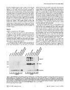
When GFP variants such as EGFP or its color variants were outside the b-can structure, could be replaced with serine without
loss of brightness. In the case of C70, site-saturated mutagenesis
expressed in the ER by the addition of the signal sequence (ss) of showed that methionine was the only amino acid that could
a1-antitrypsin and the cells then incubated in the presence of functionally substitute C70, whose thiol group is located inside the
brefeldin A to inhibit ER exit, most of the variants formed b-can. The SGFP2 variant possessing the double mutant, C48S/
covalently-linked oligomers (Figure 1, lanes 10 and 12). Even a C70M, was called cf(cysteine-free)SGFP2 (Figure 1, lane 15).
‘‘monomeric mutation’’, A206K [25], that greatly reduces subunit
association by introducing charge-repulsion at the dimer interface In contrast to the extremely low brightness of the C48S/C70S
had no effect on oligomerization (lane11). GFP mutant [20], cfSGFP2 apparently retained most of its
brightness. To quantify the specific brightness, we analyzed the
To understand whether this oligomerization was caused by the photon counting histogram (PCH). This technique was developed
intrinsic intersubunit affinity of GFP or occurred stochastically due to account for the fluctuations in fluorescence amplitude for
to the high redox-potential of the ER lumen, we examined molecules diffusing through an observation volume [27]. Here, we
Figure 1. Oligomerization of GFP family proteins in the ER. Various GFP family proteins as indicated in the figure were targeted to the ER by a
signal sequence (‘‘ss’’) and subjected to SDS-PAGE after SDS treatment at 70uC for 10 min with (lanes 1–8) or without (lanes 9–16) reduction as
described in Materials and Methods. Shown is an immunoblot probed with anti-GFP antibody. The position of each oligomer is indicated. Vehicle:
mock-transfection. *: unidentified bands. The numbers to the left indicate the positions of molecular mass markers. When molecules contained two
cysteine residues, they formed higher molecular mass oligomers (lanes 10, 11, 12 and 13) whereas molecules containing a single cysteine formed a
dimer (lanes 14 and 16). The monomer form SGFP2(C48S/C70M) (lane 15) is named cfSGFP2.
doi:10.1371/journal.pone.0037551.g001
PLoS ONE | www.plosone.org 2 May 2012 | Volume 7 | Issue 5 | e37551