
Page 27 - Human Environment Interface (3)
P. 27
Novel Fluorescent Probes for the Oxidative Milieu
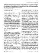
The major band of ss-cgfTagRFP (arrow) migrated faster than ss-cfTagRFP. Tunicamycin treatment (5 mg/mL) for 8 hr caused faster migration (lane 10,
‘‘aglyco’’) of ss-TagRFP (lane9, ‘‘glycol’’). In contrast, ss-cgfTagRFP showed no difference in mobility (lanes 11 and 12). Bands of unknown origin that
are inconsistently observed with ss-cgfTagRFP (lane 8) or ss-TagRFP (lanes 9 and 10) are indicated by an asterisk (*). Schematic drawings of mutations
in cgfTagRFP are also shown. (B) Excitation and emission spectra of newly developed fluorescent proteins. Normalized absorption (left) or emission
spectra (right) of the purified fluorescent proteins. (C) Localization of ss-cgfTagRFP in cells treated with brefeldin A. Fluorescence of ss-cgfTagRFP
almost completely matched with anti-calnexin antibody immunofluorescence.
doi:10.1371/journal.pone.0037551.g008
analysis of cgfmKate2 showed that the in vivo brightness was oligomers in the ER are apparently formed before obtaining a
unchanged from mKate2 (Figure 3). stable interface. This process, which is specific rather than
random, probably occurs due to intrinsic affinity (Figure 2). It is
Photochemical Properties thus logical that increasing the folding rate of GFP decreases the
To examine photochemical properties of a set of cysteine-free extent of oligomerization. It has been reported that Superfolder
GFP could escape cysteine-mediated oligomerization [23], and we
fluorescent proteins as described above, we carried out several also confirmed that SuperFast GFP formed less amounts of
measurements of the purified proteins. cfSGFP2 showed bright- covalent dimers albeit it did not eliminate formation of the
ness very similar to SGFP2 with a higher quantum yield (Table 1). oxidized dimer (Figure 1, lane 16). Since it was impossible to
As with SGFP, cfSGFP2 showed red-shift absorbance compared to identify the residues that are responsible for inter-subunit
EGFP, albeit the shift of cfSGFP2 was slightly smaller than SGFP association during folding, we instead tested the possibility that
(Figure 8B anf Table 1). It should be noted that a characteristic cysteine residues could be substituted with other amino acids.
peak of SGFP2 at 396 nm, which is not prominent in EGFP, was Although the new set of fluorescent tags described here do not
reduced by 22% in cfSGFP2. seem to hamper or slow the folding process, caution is needed
since the self-assembling property of fluorescent proteins during
In vivo molecular brightness of cgfTagRFP was comparable to folding may not be eliminated.
TagRFP (Figure 3 and Table 1) and the isolated protein also
showed similar brightness (78%) compared to TagRFP (Table 1). The surprising difference in folding and targeting of the prion
The bleaching time constant of cgfTagRFP was only slightly lower fusion protein by the presence of cysteines in the tag indicates that
than TagRFP. pKa values of both red fluorescent proteins were folding of a tag per se could have global effects on the tagged target
near 3 (Figure S3 and Table 1), indicating that the acid-resistance (Figures 6 and 7). The prion protein has been extensively studied
of the fluorophore was not lost by the elimination of cysteine. This as a cause of transmissible spongiform encephalopathies. Confor-
property was not conserved in the red-shift variant, mKate2 whose mational alteration to the b-sheet rich structure is thought to be
pKa was 5.4 [41]. In cgfmKate2, the pKa was reduced to 4.6 associated with the main pathogenic event [43]. Since it was
(Figure S3 and Table 1). We did not include the results of mKate2 beyond the scope of this study, we have not determined whether
in Table 1 since our mKate2 preparations were unstable when the SDS-resistant form of the prion fusion protein described in
isolated from bacteria. Consistent wth the comparable in vivo Figure 6 is related to the pathogenic form, which exhibits
molecular brightness of cgfmKate2 to the parent protein, the proteinase K resistance. This unusual property might have
quantum yield of cgfmKate2 (0.47) was comparable to the appeared because the SGFP2 tag containing an artificial signal
reported quantum yield (0.40) of mKate2. sequence was fused to the N-terminus of prion at K23. Several
studies have successfully demonstrated proper targeting of EGFP-
Discussion prion fusion protein to the cell surface and generation of a
transgenic mouse expressing the fusion protein when EGFP was
GFP technology has become a routine basic methodology in inserted at the N-terminus of the GPI-attachment site [44] or at
various fields of the life sciences. This has enabled analysis of the the C-terminus of the signal sequence cleavage site [45].
dynamics of molecules in the living state. For example, diffusion is
the most basic parameter of any reaction and only the Since the thiol groups of two cysteine residues in GFP family
development of GFP revealed how genetically coded molecules members are located inside the tight b-can structure, it was not
actually propagate in cells. However, usage of the GFP tag in the surprising that disulfide bonded oligomers emit no fluorescence.
secretory pathway seems to be limited. We considered that the However, why then do the cysteine residues that are supposedly
limitation of GFP technology in the secretory pathway was related protected from solvent have an effect on diffusion? One possibility
to the anomalous property of motion in the lumen of the ER and is that the oligomers caused a crowding effect on the normal
the reactivity of the cysteine residues in GFP family proteins in the diffusion of the fluorescent monomer. We think this unlikely
oxidative environment. To overcome these problems, in this because 1) the expression levels of two proteins (SGFP2 and
present study we have developed cysteine-free fluorescent tags and cfSGFP2) were comparable and 2) measurements were performed
identified a set of cysteine replacement mutants that could still at the sub-nanomolar, single molecule time regime. The second
retain the brightness of their wild type counterparts. We have possibility is that large oligomers may simply block the narrow
shown that the GFP mutant cfSGFP2 dramatically improved the lumen of the ER. Strictly speaking, we could not exclude this and,
diffusional property and inertness as a tag. To expand the indeed, the analysis using a confined diffusion model (Figure S1B)
repertoire of fluorescent tags that would be useful for analysis of may be interpreted that the freely diffusing space was reduced in
the secretory pathway or extracellular space, we further generated SGFP2. However, we think it unlikely because expression of the
cgfTagRFP and cgfmKate2 as cysteine-free (cf) variants of GFP-mCherry tandem dimer showed no punctate structures (our
TagRFP and mKate2, respectively. unpublished observation). Another possibility we think most likely
is that fluctuation of a b-can structure may externalize cysteine
In early studies aimed at improving fluorescent proteins as tags, residues so that they may react with other thiols or thiol-reacting
efforts were focused on eliminating the intrinsic oligomerization enzymes such as polypeptide disulfide isomerase (ER) or thior-
property of GFP or RFP (DsRed or eqFP578) family proteins edoxin (cytoplasm). This could happen stochastically, which would
[3,25,42]. These strategies were essentially based on creation of explain the time evolution of their diffusion. It should be noted
charge repulsion [42] and/or elimination of hydrophobic residues
at subunit interfaces [25]. As described previously [23], covalent
PLoS ONE | www.plosone.org 11 May 2012 | Volume 7 | Issue 5 | e37551