
Page 25 - Human Environment Interface (3)
P. 25
Novel Fluorescent Probes for the Oxidative Milieu
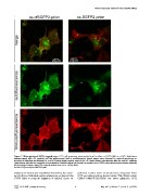
Figure 7. Mistargeting of SGFP2-tagged prion. COS7 cells expressing prion protein fused to either ss-cfSGFP2 (left) or ss-SGFP2 (right) were
immunostained with GFP antibody and the epifluorescent (red) or autofluorescent (green) images were observed by confocal microscopy as
described in Materials and Methods. In ss-cfSGFP2-prion fusion protein, most of the GFP signal showed colocalization with the anti-GFP antibody
signal (merge, left) with the exception of a perinuclear, Golgi-like region (see Results). In contrast, the ss-SGFP2-prion fusion protein showed markedly
distinct images (merge, right). The confocal pinhole was set to 1.0 Airy Units.
doi:10.1371/journal.pone.0037551.g007
brightness of mKate2 (our unpublished observation). By examin- performed another round of site-saturated mutagenesis where
ing the effects of individual cysteine substitutions, we observed that C172 was replaced with an alanine residue. This mKate2 variant
C172V failed to retain the brightness of mKate2. Hence we C26A/C114M/C172A/C222S was called cgfmKate2. PCH
PLoS ONE | www.plosone.org 9 May 2012 | Volume 7 | Issue 5 | e37551