
Page 58 - Human Environment Interface (3)
P. 58
Brain Barrier Integrity and Development in the Rat
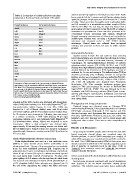
Table 3. Comparison of cellular adhesion transcripts solution was directly applied to choroid plexus tissue of the fourth
expressed at the blood-brain and blood-CSF barriers. brain ventricle, left for 5 minutes and cold fixative solution slowly
applied to plexuses. All plexuses were left in fixative for 2–3 hours
Present at plexus Not present at plexus in fridge. Tissues were then thoroughly rinsed in phosphate buffer
and left overnight in a streptavidin-peroxidase complex (Vector
Amotl2 Amot ABC kit PK-6100). The next day, the tissue was stained with a
Cdh5 Amotl1 nickel-enhanced DAB reaction (Vector kit SK-4100) according to
Cgnl1 Arhgap17 manufacturer’s specifications. Tissue was then processed as for
Cldn12 Ash1l conventional electron microscopy with osmium, dehydrated
Dlg1 Cldn5 through increasing concentrations of ethanol and embedded in
Dlgap1 Jam4 araldite–epon. Sections were cut using a Reichard Ultracut E
Esam Mpp1 microtome and examined under a LEO 912AB electron
F11r Tjap microscope. Control tissue was collected from un-injected
Jam2 Tjp2 embryos, was processed as above but gave no visible reaction
Lin7c Wnk1 product.
Magi3
Marveld2 Immunohistochemistry
Mpp5 Sagittal sections through E15 and adult rat brain including
Mpp7
Ocln lateral choroid plexus were selected from the collection of rat tissue
Pard3 at the Faculty of Health and Medical Sciences, University of
Pard6g Copenhagen, for immunohistochemical detection of carbonic
Scrib anhydrase-related protein VIII (CA8), SLC4A1 and CLIC3.
Tjp1 Sections were deparaffinised in xylene, rehydrated through graded
alcohols followed by treatments in 0.5% hydrogen peroxide in
Comparison of data presented in the current study on blood-CSF barrier methanol for 15 min and rinsing in Tris-buffered saline (TBS) as
transcriptome and the previously published blood-brain barrier transcriptome described previously [13]. Following removal of non-specific
[33]. Most (19 of 29) junction transcripts enriched at the blood-brain barrier binding, sections were incubated in primary antibodies (SLC4A1,
were present at the blood-CSF barrier. Only one transcript thought to be brain- 8566-1-AP, 1:400; CLIC315971-1-AP, 1:200; or CA8, 12391-1-
barrier specific (Jam4) was not present at the blood-CSF barrier. Note that our AP, 1:150. All antibodies were obtained from Proteintech,
data are from E15 rat choroid plexus, whereas blood-brain barrier [33] are from Manchester, UK). After overnight incubation sections were
P2-8 mouse cerebral endothelial cells. washed in TBS and incubated for 30 min in EnVisionTM+
doi:10.1371/journal.pone.0065629.t003 System/HRP (DAKO), K5007. This was followed by 6 min
incubation with DAB-chromogen solution (DAKO) and counter-
minutes) and the ABC reaction was developed with diaminoben- staining with Mayer’s haematoxylin, dehydrated and mounted
zidine (DAB)/nickel staining. One DAB tablet (SigmaFastTM 3,39- with DPX. Control sections contained no primary antibodies and
were always blank.
diaminobenzidine tablets, Sigma, St Lois, MO, USA) was
dissolved in 1 ml of filtered distilled water with 40 ml nickel Photography and image preparation
Digitized images were obtained using an Olympus DP70
ammonium sulphate, with the final solution used to incubate tissue
camera housing (Olympus, Tokyo, Japan) attached to an Olympus
for 10 minutes at room temperature. Following, tissue was moved BX50 light microscope (Olympus). A 106 eyepiece and 406
to a solution containing 1 DAB tablet (Sigma), 40 ml nickel objective lens were used. Raw image files were process in Adobe
ammonium sulphate and 1 urea hydroxide tablet (SigmaFastTM Photoshop CS5H (Adobe H Systems, San Jose, CA, USA). The
3,39-diaminobenzidine tablets, Sigma) and incubated at room brightness and curve functions were used to obtain images with
background close to white. There was no other manipulation of
temperature. When a dark precipitate was observed (after images.
approximately 10 minutes) the staining reaction was halted with Results and Discussion
filtered distilled water. Stained sections were mounted on silanized Sequencing of total RNA samples collected from E15 and adult
lateral ventricular choroid plexus was completed using the
glass slides with fluorescent mounting media (DAKO) and dried at Illumina platform. A total of 29479 transcripts were mapped, of
4uC overnight. which 5872 transcripts were enriched at E15 and 4781 enriched in
the adult. Over 18000 (approximately 60%) of these genes were
Biotin ethylenediamine, electron microscopy already expressed at adult levels in E15 samples, suggesting a great
Biotin-ethylenediamine was used as a low molecular tracer deal of maturity of expression levels in this tissue. Refer to
Table S1 (raw transcript count) and Table S2 (summarised
(286Da) to assess choroid plexus barrier permeability at an analysed data) for full dataset.
ultrastructural level. The use of this compound as a permeability
tracer has been established previously [24]. Pregnant rats at Genes associated with choroid plexus differentiation and
gestational day 15 were anaesthetised and embryos externalized. development
About 0.5 mL of tracer (0.2 mg/mL in saline) was slowly injected
into each lateral brain ventricle of embryos (n = 4) using a fine Several genes previously associated with neural development
glass capillary. At 5–8 minutes after the injection, the whole brain were detected in the rat lateral ventricular choroid plexus
was removed and submerged in fixative (2.5% glutaraldehyde in epithelium. This included many transcription factors traditionally
pH7.4 phosphate buffer), before plexuses were dissected out. At
the end of the experiment, the mother was killed and tracer
PLOS ONE | www.plosone.org 5 July 2013 | Volume 8 | Issue 7 | e65629