
Page 67 - Human Environment Interface (3)
P. 67
Brain Barrier Integrity and Development in the Rat
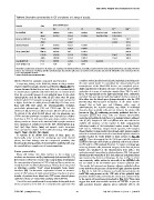
Table 6. Electrolyte concentrations in CSF and plasma of a range of species.
Species Age Ions (CSF/Plasma)
Rat (21d)[82]
Monkey (168d) [63] NB Na+ K+ Cl2 HCO32 Ca2+ Mg2+
Rabbit (28d) [82] Adult
Sheep (150d) [64] E50-60 144/150 3.9/6.5 108/102 24.3/25.5 [114] 2.4/2.9 [115] -
Adult 152/153 3.2/5.0 125/105 25.8/20.5 [116] 1.4/2.5 [115] -
Dog (62d) [112] P20 - 3.6/4.5 - - 3.8/4.6 1.9/1.2
Horse (340d) [113] Adult - 2.6/4.5 - - 2.3/4.6 1.9/1.2
E44-50 143/143 4.1/3.1 113/102 - - 1.5/3.2
E85-90 152/155 3.0/3.2 120/102 - - 1.5/2.2
Adult 135/138 5.4/- 113/113 - - 2.0/1.6
P1-4 144/136 3.5/3.8 123/104 - 3.4/6.1 1.9/1.8
P1-2 148/138 3.1/3.4 128/115 - 2.5/3.4 1.8/1.4
149/141 3.3/4.3 122/104 26.5/25.6 - -
143/- 3.6/- 109/- - - -
Electrolyte concentrations (mequiv/L or kg H20) in CSF and plasma of embryos/newborns and adults. Gestation term for each species listed in days in brackets. Note that
there is net movements of sodium, chloride and bicarbonate ions into the CSF, and a net movement of potassium ions into the blood. Net water movement into the
CSF is by aquaporin channels.
doi:10.1371/journal.pone.0065629.t006
Genes related to synaptic structure and function combines molecular (biochemical) and physiological techniques in
A surprising finding in the RNA-Seq dataset is a large number the same animal model. To consolidate the data provided by the
transcriptome of the blood-CSF barrier, we completed perme-
of genes related to synaptic structure and function (Table S5). For ability experiments to illustrate the route of entry for water-soluble
reasons discussed below they are very likely to be expressed solely molecules of a range of molecular weights (from 286 Da and 3
in choroid plexus epithelia cells rather than due to contamination kDa). For experiments at the light microscopic level, embryonic
from the very small amount of non-epithelial tissue in the whole and adult rats were injected with biotinylated dextran amines of 3
choroid plexus used for this study. At E15 there were 64 genes kDa either into the blood space (intraperitoneal) or into the CSF
defined by their GO categories as synapse-related and expressed at (intrathecally). Microscopical localisation of the biotin marker
a higher level than in adult plexus (Table S5). Of these, more showed that at both ages and following either route of
than half (28) were genes for neurotransmitter receptors, administration, the reaction product was visible in individual
particularly glutamatergic (14) and GABA-ergic (5) but also choroid plexus epithelial cells and not between the plexus cells
cholinergic (4), serotonergic (2) purinergic (2) and adrenergic (1). (arrows in Fig. 4) confirming previously published data for a
In contrast in the adult choroid plexus only one glutamate, two marsupial species that these cells can transfer water-soluble
GABA and two purinergic receptors were expressed at a higher molecules intracellularly, but there is no leakage between adjacent
level than at E15. At both ages there was a large number of genes cells [17,47]. Samples of CSF and plasma were also collected to
whose proteins are known to be associated with synaptic structures confirm the presence of the marker in both compartments
were expressed at a higher level in the E15 choroid plexus (e.g. indicating transfer of the biotinylated marker across the blood-
Syn1, Syn3, Nrgn, Stx1za, Synpo), and synaptic vesicle transcripts CSF barrier. Following intraperitoneal injection, CSF samples
were expressed at a higher level in adult plexus (e.g. Sv2b, Syngr1, showed biotin reaction product (not shown), while plasma samples
Snap25, Vamp1, Syp Syt1, Syt4, Snapin). taken from animals that received an intrathecal injection showed a
positive reaction for biotin (visible in tissue sections in Fig. 4B
It remains to be shown how many of these genes are and D). These data suggest that the 3 kDa water soluble marker is
functionally active in both the developing and adult choroid transferred through choroid plexus epithelial cells in both a blood-
plexus, but these findings raise the unexpected possibility that to-CSF and in a CSF-to-blood direction. To ensure no leakage was
functional (secretory) activity of choroid plexus epithelial cells may occurring between cells, functional integrity of the choroid plexus
be controlled by a complex set of neurotransmitters. barrier was visualised at the ultrastructural level with a low
molecular weight tracer (biotin ethylenediamine, 286 Da) injected
Barrier permeability directly into the ventricles of E15 and adult rats and plexus tissue
Many still believe that brain barriers in the embryo and was examined by electron microscopy. As seen in Fig. 5, tracer
was extensively visible within the microvilli of the epithelial cells
newborn are immature and (by implication) dysfunctional [85–87] but not visible in the intercellular space between epithelial cells.
(for review see [6]). One reason is that early in brain development The marker did not penetrate between adjacent epithelial cells as
the concentration of proteins in CSF is high and these proteins are it was halted by the presence of functional tight junctions (arrows
mostly derived from blood plasma [5,6]. This has been interpreted in Fig. 5). In both the embryonic and adult choroid plexus the
as reflecting a passive ‘‘leak’’ across incomplete brain barrier tracer was only present on the CSF side of the epithelial tight
interfaces [88]. An alternative explanation is based on measure- junctions. Small vesicles containing the tracer were common close
ments of CSF volume of distribution [9,17] and evidence of active to the apical cell membrane (arrowhead in Fig. 5C). These data
transfer of proteins from blood into CSF across choroid plexus suggest that small water-soluble markers are not able to pass the
epithelial cells by an intracellular pathway [10,17,89–93]. tight junctions between adjacent plexus epithelial cells in the
One of the main problems with many studies dealing with brain
barrier properties is the lack of a comprehensive approach that
PLOS ONE | www.plosone.org 14 July 2013 | Volume 8 | Issue 7 | e65629