
Page 69 - Human Environment Interface (3)
P. 69
Brain Barrier Integrity and Development in the Rat
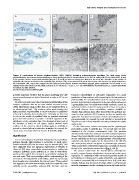
Figure 5. Localisation of biotin ethylenediamine (BED, 286Da) following intraventricular injection. The small tracer, biotin
ethylenediamine, was injected intraventricularly to assess barrier function in choroid plexus in the E15 rat embryo (A–C) and adult (D–F). A and
D are semithin sections stained with toluidine blue; B, C, E and F are electron micrographs. Note that the tracer was abundant on the outside of
epithelial cells but was restricted by the epithelial tight junctions (TJ), present towards the apical side of these cells and was not visible further into
the intercellular cleft. These data suggest that the tight junctions present between intimately apposed plexus epithelium are functional in E15
animals to the same extent as in the adult. Scale bars: A, D = 50 mm, B, E = 5 mm, C, F = 1 mm. Abbreviations: BV, blood vessel; CSF, cerebrospinal fluid;
N, nucleus; TJ, tight junction.
doi:10.1371/journal.pone.0065629.g005
provides important evidence that the genes underlying key CSF Diamond acknowledged an alternative explanation of a small
secretory mechanisms are indeed functional as early as E15 in rat population of low resistance cells accounting for the low resistance
embryos. pathway [98]. An overall problem with the concept of the tight
junction and associated components of the paracellular pathway as
In order to provide some direct functional understanding of this a permeability route for small water-soluble molecules across an
complex expression data set we have carried out some perme- epithelial interface is that for many years there have been only a
ability experiments using markers that can be visualised down to few small electron dense extracellular markers available to
an ultrastructural level. This confirms earlier work showing that visualize this pathway; these often showed that this pathway was
tight junctional impermeability to small molecules is established closed to these molecules [99]. However, as they are usually only
very early in choroid plexus development. Otherwise we have had applied for very short times because of their toxicity (lanthanum or
to rely on the wealth of published data on junctions associated pyroantimonate), for example by local vascular or parenchymal
genes and their proteins to provide a detailed appraisal of the injection or even in fixed materials it is unclear whether the results
significance of the expression data. One drawback is that much of apply in vivo.
the literature is based on cell culture studies, without much
evidence of the extent to which findings apply to epithelia in On the other hand, markers commonly used for physiological
general or choroid plexus in particular, in vivo (see for example the permeability studies in epithelia (e.g. 14C-sucrose) have not been
recent series of articles edited by Fromm & Schulzke [94]). visualised at the electron microscopical level. Our previous
ultrastructural results from studies of neonatal marsupial opossums
Significance showed that biotin-labelled small molecular sized surrogates for
sucrose and inulin are seen in a small proportion of choroid plexus
It is a central dogma of epithelial biology that the paracellular epithelial cells, whether applied on the blood [47] or the CSF [9]
pathway through tight junctions is permeable to small water- side of the plexus, and do not appear to permeate tight junctions.
soluble molecules [95] but the extent may be variable [96]. This This has now been confirmed for embryonic and adult rat choroid
view originated from the ingenious experiments of Fro¨mter & plexus in the experiments reported here (Fig. 4 and 5).
Diamond [97] using extracellular microelectrodes to localise low
resistance pathways across epithelia at or close to intercellular The present results from the choroid plexus transcriptome
junctions from which they proposed a paracellular pathway for provide a comprehensive identification of junctional protein gene
ions and water. This was a paradigm shift in understanding of the expression. In combination with current and previous studies of
mechanism of transepithelial permeability, which had previously permeability in embryos and in adults [5,9,47,91] and supported
been thought to be transcellular for ions and water. These authors by results in postnatal [33] and embryonic mice [13], these data
also suggested that small water-soluble molecules might also cross strongly indicate that the brain develops within a well-protected
epithelia via the paracellular pathway [97], but later work by internal environment and the exchange between the brain and the
PLOS ONE | www.plosone.org 16 July 2013 | Volume 8 | Issue 7 | e65629