
Page 27 - Human Environment Interface (4)
P. 27
Surface Chemistry Directs Protein Remodeling
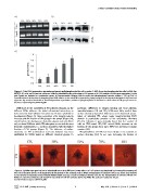
Figure 4. Total FAK expression (protein and gene) and phosphorylation of tyrosine Y-397, the autophosphorylation site in FAK, for
MC3T3-E1 cells on FN coated surfaces. SAMs are identified by the percentage of OH groups. a) RT-PCR analysis of FAKs gene expression, b-actin
and Gapdh are included as constitutive genes. b) Representative Western blot for total and phophorylated tyrosine residue Y-397 on FAK. c)
Quantification of the fraction of phosphorylated FAKs relative to the total FAK expression by image analysis of the western blot bands in b). Error bars
represent the standard deviation of three independent experiments; enhanced phosphorylation is obtained as the fraction of OH groups increases.
doi:10.1371/journal.pone.0019610.g004
Differences in the availability of FN adhesion domains on the particular, differences in integrin binding and focal adhesion
different SAMs influence the initial cell-material interaction, as assembly between OH and CH3 SAMs most likely resulted from
determined by focal adhesion formation and F-actin cytoskeleton surface chemistry dependent differences in the functional presen-
development (Figure 3). Gene expression of b1 integrin subunit tation of adsorbed FN, whose major integrin-binding RGD
increases with the fraction of OH groups in the sample (Figure S4), domain is particularly sensitive to the underlying chemistry
which leads to the development of vinculin plaques and actin fibers [41,45]. Likewise, it was previously found that the number of
only on those SAMs on which FN adsorption occurs with the most cells on FBS-coated CH3/OH mixed SAMs increases as the
favorable conformation, i.e. on those chemistries with the highest fraction of OH groups does; up to 80% OH and then it remains
fraction of OH groups (Figure 3). The influence of surface constant [46].
chemistry on FN conformation and cell adhesion has been
established for SAMs based on different chemical groups. In Phosphorylation of FAK has been shown to be sensitive to
surface chemistry [45]. In our case, increasing the fraction of
Figure 5. Cellular reorganization of adsorbed FN on the different SAMs after 2.5 h of culture as obtained by immunofluorecence of
FN. The red bottom shows FN homogeneously distributed on the material surface. When reorganization of adsorbed FN occurs, black areas (related
to the removal of substrate-bound FN) and fibrillar bright areas (as a result of enhanced fluorescence for the incorporation of removed FN into FN-
fibrils) are observed. Only the cell shadow in observed for low OH contents (CH3 and 30%). The scale bar represents 50 mm.
doi:10.1371/journal.pone.0019610.g005
PLoS ONE | www.plosone.org 5 May 2011 | Volume 6 | Issue 5 | e19610