
Page 60 - Human Environment Interface (4)
P. 60
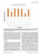
Epigenetics of the Placenta

Figure 7. Real time PCR analysis was performed using total RNA samples from MBC (n = 6) and CVS (n = 6) to detect transcript levels
encoded by the following genes: COL15A1, GJA1, LAMB1, LUM, PITX2, SLC16A4, TFPI2 and VGLL3. Each reaction was run in triplicate
against an endogenous control (GUSB) and normalized against one of the MBC samples. The median logRQ was plotted for each gene.
doi:10.1371/journal.pone.0014723.g007

pattern, whereas when CpG frequency is . 0.4, for genes with                bimodal distribution of hyper- and hypomethylated sites. We found
hypomethylated promoters, gene expression is positively correlat-           these to be global phenomena, for which the biological basis is
ed with hypomethylation level, or equivalently, negatively                  unclear. These observations may however be related to the fact that
correlated with methylation level (Figure 9D). The data presented           very early gestational trophoblast stem cells display a hypomethy-
in Figure 9D are consistent with our previous observations of the           lated genomic state that is consistent with a semi-pluripotent
relationship between promoter methylation and gene expression               phenotype[35]. This is supported by the fact that trophoblast
(without considering CpG frequency) based on data obtained                  lineages are thought to retain pluripotency for some time after
using both the Agilent array and Infinium arrays (Figure 8A,B,C).           implantation[36] and the observation that relative hypomethylation
That is, promoter methylation level is negatively correlated with           in CVS versus MBC in the first trimester is lost by the third
gene expression, especially for genes with relatively hypomethy-            trimester[37]. The relative hypomethylation of CVS versus MBC
lated promoter.                                                             may also be related to the highly proliferative and invasive nature of
                                                                            this tissue and its requirement for a highly active and complex
Discussion                                                                  transcriptional state. Interestingly, Papageorgiou et al., (2009)[37]
                                                                            recently reported similar findings using an immunoprecipitation-
   We present a comprehensive epigenetic analysis of the placental          based approach although this was not as pronounced as in our data
chorionic villus (CVS) and gestational age matched maternal                 and was not the case for all chromosomes.
blood cells (MBC) at the level of DNA methylation. In addition to
providing detailed insight into the structure and organization of              The spatial association of broadly hypomethylated regions
the CVS and MBC methylomes in the context of promoters, CpG                 observed in CVS and MBC genomes is intriguing. It is conceivable
islands and gene bodies, we present novel findings relating the             that such differentially methylated regions play a role in the
methylation levels of these genetic elements to gene expression             regulation of expression of functionally related genes such as those
levels, biological function and primary DNA sequence.                       identified in Table 1. The fact that some of these genes are
                                                                            aberrantly methylated at the DNA level in a variety of tumors[37]
   One fundamental difference between the CVS and MBC                       is notable given the previously noted link between the ‘‘molecular
genomes is the bias towards a hypomethylated state in the former.           phenotype’’ of tumors and early mammalian development[25].
Related to this is that fact that, unlike the differentiated adult tissues
and tumor samples we investigated, CVS genomes do not have a                   The discovery that T-DMRs are more likely to be located outside
                                                                            promoters and gene bodies suggests that tissue specific differences in

PLoS ONE | www.plosone.org                                                  12 February 2011 | Volume 6 | Issue 2 | e14723