Page 264 - First Aid for the USMLE Step 1 2020, Thirtieth edition [MedicalBooksVN.com]_Neat
P. 264

220         SECtIoN II    Pathology  ` PATHOLOGY—neOPLASIA                                                                                                                  Pathology  ` PATHOLOGY—neOPLASIA


               Tumor nomenclature    Carcinoma implies epithelial origin, whereas sarcoma denotes mesenchymal origin. Both terms
                                       generally imply malignancy.
                                     Benign tumors are usually well-differentiated and well-demarcated, with low mitotic activity, no
                                       metastases, and no necrosis.
                                     Malignant tumors (cancers) may show poor differentiation, erratic growth, local invasion,
                                       metastasis, and  apoptosis.
                                     Terms for non-neoplastic malformations include hamartoma (disorganized overgrowth of tissues in
                                       their native location, eg, Peutz-Jeghers polyps) and choristoma (normal tissue in a foreign location,
                                       eg, gastric tissue located in distal ileum in Meckel diverticulum).
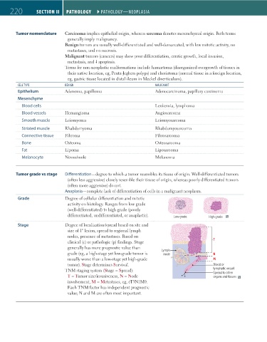
                CeLL TYPe            BenIGn                                    MALIGnAnT
                Epithelium           Adenoma, papilloma                        Adenocarcinoma, papillary carcinoma

                Mesenchyme
                  Blood cells                                                  Leukemia, lymphoma
                  Blood vessels      Hemangioma                                Angiosarcoma
                  Smooth muscle      Leiomyoma                                 Leiomyosarcoma
                  Striated muscle    Rhabdomyoma                               Rhabdomyosarcoma
                  Connective tissue  Fibroma                                   Fibrosarcoma
                  Bone               Osteoma                                   Osteosarcoma
                  Fat                Lipoma                                    Liposarcoma
                  Melanocyte         Nevus/mole                                Melanoma


               Tumor grade vs stage  Differentiation—degree to which a tumor resembles its tissue of origin. Well-differentiated tumors
                                       (often less aggressive) closely resemble their tissue of origin, whereas poorly differentiated tumors
                                       (often more aggressive) do not.
                                     Anaplasia—complete lack of differentiation of cells in a malignant neoplasm.
                Grade                Degree of cellular differentiation and mitotic
                                       activity on histology. Ranges from low grade
                                       (well-differentiated) to high grade (poorly     Low grade       High grade
                                       differentiated, undifferentiated, or anaplastic).  Low grade    High grade

                Stage                Degree of localization/spread based on site and
                                       size of 1° lesion, spread to regional lymph
                                       nodes, presence of metastases. Based on
                                       clinical (c) or pathologic (p) findings. Stage                    T
                                       generally has more prognostic value than                           T
                                       grade (eg, a high-stage yet low-grade tumor is   Lymph            N
                                                                                   node
                                                                                  Lymph
                                       usually worse than a low-stage yet high-grade   node               N M
                                       tumor). Stage determines Survival.                                 M Blood or
                                     TNM staging system (Stage = Spread):                                lymphatic vessel
                                                                                                          Blood or
                                                                                                         Spread to other
                                                                                                          lymphatic vessel
                                       T = Tumor size/invasiveness, N = Node                             organs and tissues
                                                                                                          Spread to other
                                       involvement, M = Metastases, eg, cT3N1M0.                          organs and tissues
                                       Each TNM factor has independent prognostic
                                       value; N and M are often most important.


          FAS1_2019_04-Pathol.indd   220                                                                                11/7/19   4:02 PM
   259   260   261   262   263   264   265   266   267   268   269