
Page 466 - First Aid for the USMLE Step 1 2020, Thirtieth edition [MedicalBooksVN.com]_Neat
P. 466
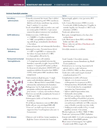
422 SectIon III Hematology and oncology ` hematology and oncology—Pathology Hematology and oncology ` hematology and oncology—Pathology
Intrinsic hemolytic anemias
deScRIPtIon FIndIngS
Hereditary Primarily autosomal dominant. Due to defect Splenomegaly, aplastic crisis (parvovirus B19
spherocytosis in proteins interacting with RBC membrane infection).
skeleton and plasma membrane (eg, ankyrin, Labs: mean fluorescence of RBCs in eosin
band 3, protein 4.2, spectrin). 5-maleimide (EMA) binding test, fragility in
Small, round RBCs with less surface area and osmotic fragility test. Normal to MCV with
no central pallor ( MCHC) premature abundance of RBCs.
removal by spleen (extravascular hemolysis). Treatment: splenectomy.
G6PD deficiency X-linked recessive. G6PD defect Back pain, hemoglobinuria a few days after
NADPH reduced glutathione oxidant stress.
RBC susceptibility to oxidative stress Labs: blood smear shows RBCs with Heinz
(eg, sulfa drugs, antimalarials, fava beans) bodies and bite cells.
hemolysis. “Stress makes me eat bites of fava beans with
Causes extravascular and intravascular hemolysis. Heinz ketchup.”
Pyruvate kinase Autosomal recessive. Pyruvate kinase defect Hemolytic anemia in a newborn.
deficiency ATP rigid RBCs extravascular
hemolysis. Increases levels of 2,3-BPG
hemoglobin affinity for O .
2
Paroxysmal nocturnal Hematopoietic stem cell mutation Triad: Coombs ⊝ hemolytic anemia,
hemoglobinuria complement-mediated intravascular pancytopenia, venous thrombosis (eg, Budd-
hemolysis, especially at night. Acquired PIGA Chiari syndrome).
mutation impaired GPI anchor synthesis Pink/red urine in morning. Associated with
for decay-accelerating factor (DAF/CD55) and aplastic anemia, acute leukemias.
membrane inhibitor of reactive lysis (MIRL/ Labs: CD55/59 ⊝ RBCs on flow cytometry.
CD59), which protect RBC membrane from Treatment: eculizumab (targets terminal
complement. complement protein C5).
Sickle cell anemia Point mutation in β-globin gene single Complications in sickle cell disease:
A amino acid substitution (glutamic acid Aplastic crisis (transient arrest of
valine). Mutant HbA is termed HbS. Causes erythropoiesis due to parvovirus B19).
extravascular and intravascular hemolysis. Autosplenectomy (Howell-Jolly bodies)
Pathogenesis: low O , high altitude, or acidosis risk of infection by encapsulated
2
precipitates sickling (deoxygenated HbS organisms (eg, S pneumoniae).
polymerizes) anemia, vaso-occlusive disease. Splenic infarct/sequestration crisis.
Newborns are initially asymptomatic because of Salmonella osteomyelitis.
HbF and HbS. Painful vaso-occlusive crises: dactylitis
Heterozygotes (sickle cell trait) have resistance (painful swelling of hands/feet), priapism,
to malaria. acute chest syndrome (respiratory distress,
8% of African Americans carry an HbS allele. new pulmonary infiltrates on CXR, common
Sickle cells are crescent-shaped RBCs A . cause of death), avascular necrosis, stroke.
“Crew cut” on skull x-ray due to marrow Sickling in renal medulla ( Po ) renal
2
expansion from erythropoiesis (also seen in papillary necrosis hematuria.
thalassemias). Hb electrophoresis: HbA, HbF, HbS.
Treatment: hydroxyurea ( HbF), hydration.
HbC disease Glutamic acid–to-lyCine (lysine) mutation in Patients with HbSC (1 of each mutant gene) have
β-globin. Causes extravascular hemolysis. milder disease than HbSS patients.
Blood smear in homozygotes: hemoglobin
Crystals inside RBCs, target cells.
FAS1_2019_10-HemaOncol.indd 422 11/7/19 5:05 PM