
Page 82 - Basic Principles of Textile Coloration
P. 82
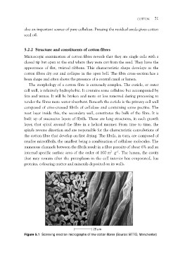
COTTON 71

also an important source of pure cellulose. Pressing the residual seeds gives cotton
seed oil.

5.2.2 Structure and constituents of cotton fibres
Microscopic examination of cotton fibres reveals that they are single cells with a
closed tip but open at the end where they were cut from the seed. They have the
appearance of flat, twisted ribbons. This characteristic shape develops as the
cotton fibres dry out and collapse in the open boll. The fibre cross-section has a
bean shape and often shows the presence of a central canal or lumen.

   The morphology of a cotton fibre is extremely complex. The cuticle, or outer
cell wall, is relatively hydrophobic. It contains some cellulose but accompanied by
fats and waxes. It will be broken and more or less removed during processing to
render the fibres more water-absorbent. Beneath the cuticle is the primary cell wall
composed of criss-crossed fibrils of cellulose and containing some pectins. The
next layer inside this, the secondary wall, constitutes the bulk of the fibre. It is
built up of successive layers of fibrils. These are long structures, in each growth
layer, that spiral around the fibre in a helical manner. From time to time, the
spirals reverse direction and are responsible for the characteristic convolutions of
the cotton fibre that develop on first drying. The fibrils, in turn, are composed of
smaller microfibrils, the smallest being a combination of cellulose molecules. The
numerous channels between the fibrils result in a fibre porosity of about 6% and an
internal specific surface area of the order of 100 m2 g–1. The lumen, the cavity
that may remain after the protoplasm in the cell interior has evaporated, has
proteins, colouring matter and minerals deposited on its walls.

                                                                          25 mm
Figure 5.1 Scanning electron micrographs of raw cotton fibres (Source: BTTG, Manchester)