
Page 365 - fbkCardioDiabetes_2017
P. 365
Cardio Diabetes Medicine 2017 341
shaped deep T wave inversions in anterior and lateral
leads suggest HOCM.
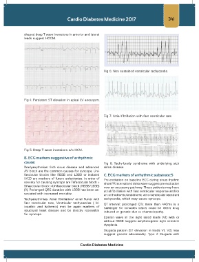
Fig 6: Non sustained ventricular tachycardia.
Fig 4: Persistent ST elevation in apical LV aneurysm.
Fig 7: Atrial fibrillation with fast ventricular rate.
Fig 5: Deep T wave inversions s/o HCM.
B. ECG markers suggestive of arrhythmic
cause: Fig 8: Tachy-brady syndrome with underlying sick
Bradyarrythmias: Sick sinus disease and advanced sinus disease.
AV block are the common causes for syncope. Uni-
fascicular blocks like RBBB and LBBB or isolated C. ECG markers of arrhythmic substrate:S
IVCD are markers of future arrhythmias. In order of Pre-excitation on baseline ECG during sinus rhythm:
severity for causing syncope are Trifascicular block > short PR interval and delta wave suggest pre excitation
Bifascicular block >Unifascicular block (RBBB/LBBB) over an accessory pathway. These patients may have
(9). Prolonged QRS duration with LBBB has been as- atrial fibrillation with fast ventricular response and/or
sociated with increased mortality.
an orthodromic/antidromic atrio-ventricular reentrant
Tachyarrythmias: Atrial fibrillation/ atrial flutter with tachycardia, which may cause syncope.
fast ventricular rate, Ventricular tachycardias ( Id- QT interval: prolonged QTc more than 440ms is a
iopathic and Ischemic) may be again markers of harbinger for torsades which could be either drug
structural heart disease and be directly reponsible induced or genetic due to channelopathy.
for syncope.
Epsilon wave in the right sided leads (V1) with or
without RBBB suggest arrythmogenic right ventricle
dysplasia.
Brugada pattern (ST elevation in leads V1, V2) may
suggest genetic abnormality. Type 2 Brugada with
Cardio Diabetes Medicine