
Page 145 - Color_Atlas_of_Physiology_5th_Ed._-_A._Despopoulos_2003
P. 145
+
Respiratory Control and Stimulation stronger when P CO 2 and/or the H concentra-
tion in blood also increase.
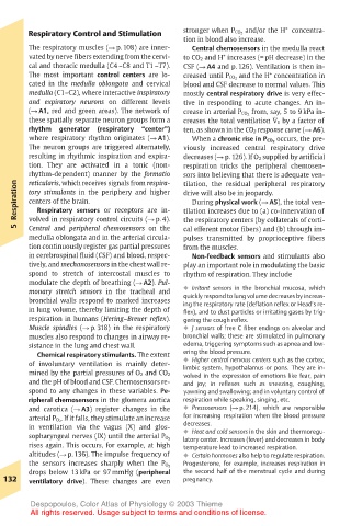
The respiratory muscles (! p. 108) are inner- Central chemosensors in the medulla react
+
vated by nerve fibers extending from the cervi- to CO 2 and H increases (= pH decrease) in the
cal and thoracic medulla (C4 –C8 and T1 –T7). CSF (! A4 and p. 126). Ventilation is then in-
The most important control centers are lo- creased until P CO 2 and the H concentration in
+
cated in the medulla oblongata and cervical blood and CSF decrease to normal values. This
medulla (C1–C2), where interactive inspiratory mostly central respiratory drive is very effec-
and expiratory neurons on different levels tive in responding to acute changes. An in-
(! A1, red and green areas). The network of crease in arterial P CO 2 from, say, 5 to 9 kPa in-
.
these spatially separate neuron groups form a creases the total ventilation V E by a factor of
rhythm generator (respiratory “center”) ten, as shown in the CO 2 response curve (! A6).
where respiratory rhythm originates (! A1). When a chronic rise in P CO 2 occurs, the pre-
The neuron groups are triggered alternately, viously increased central respiratory drive
resulting in rhythmic inspiration and expira- decreases (! p. 126). If O 2 supplied by artificial
tion. They are activated in a tonic (non- respiration tricks the peripheral chemosen-
rhythm-dependent) manner by the formatio sors into believing that there is adequate ven-
reticularis, which receives signals from respira-
Respiration tory stimulants in the periphery and higher drive will also be in jeopardy.
tilation, the residual peripheral respiratory
During physical work (! A5), the total ven-
centers of the brain.
Respiratory sensors or receptors are in-
tilation increases due to (a) co-innervation of
volved in respiratory control circuits (! p. 4).
5 Central and peripheral chemosensors on the the respiratory centers (by collaterals of corti-
cal efferent motor fibers) and (b) through im-
medulla oblongata and in the arterial circula- pulses transmitted by proprioceptive fibers
tion continuously register gas partial pressures from the muscles.
in cerebrospinal fluid (CSF) and blood, respec- Non-feedback sensors and stimulants also
tively, and mechanosensors in the chest wall re- play an important role in modulating the basic
spond to stretch of intercostal muscles to rhythm of respiration. They include
modulate the depth of breathing (! A2). Pul-
monary stretch sensors in the tracheal and ! Irritant sensors in the bronchial mucosa, which
quickly respond to lung volume decreases by increas-
bronchial walls respond to marked increases ing the respiratory rate (deflation reflex or Head’s re-
in lung volume, thereby limiting the depth of flex), and to dust particles or irritating gases by trig-
respiration in humans (Hering–Breuer reflex). gering the cough reflex.
Muscle spindles (! p. 318) in the respiratory ! J sensors of free C fiber endings on alveolar and
muscles also respond to changes in airway re- bronchial walls; these are stimulated in pulmonary
sistance in the lung and chest wall. edema, triggering symptoms such as apnea and low-
Chemical respiratory stimulants. The extent ering the blood pressure.
of involuntary ventilation is mainly deter- ! Higher central nervous centers such as the cortex,
limbic system, hypothalamus or pons. They are in-
mined by the partial pressures of O 2 and CO 2 volved in the expression of emotions like fear, pain
and the pH of blood and CSF. Chemosensors re- and joy; in reflexes such as sneezing, coughing,
spond to any changes in these variables. Pe- yawning and swallowing; and in voluntary control of
ripheral chemosensors in the glomera aortica respiration while speaking, singing, etc.
and carotica (! A3) register changes in the ! Pressosensors (! p. 214), which are responsible
arterial P O 2 . If it falls, they stimulate an increase for increasing respiration when the blood pressure
in ventilation via the vagus (X) and glos- decreases.
! Heat and cold sensors in the skin and thermoregu-
sopharyngeal nerves (IX) until the arterial P O 2 latory center. Increases (fever) and decreases in body
rises again. This occurs, for example, at high temperature lead to increased respiration.
altitudes (! p. 136). The impulse frequency of ! Certain hormones also help to regulate respiration.
Progesterone, for example, increases respiration in
the sensors increases sharply when the P O 2
drops below 13 kPa or 97 mmHg (peripheral the second half of the menstrual cycle and during
132 ventilatory drive). These changes are even pregnancy.
Despopoulos, Color Atlas of Physiology © 2003 Thieme
All rights reserved. Usage subject to terms and conditions of license.