
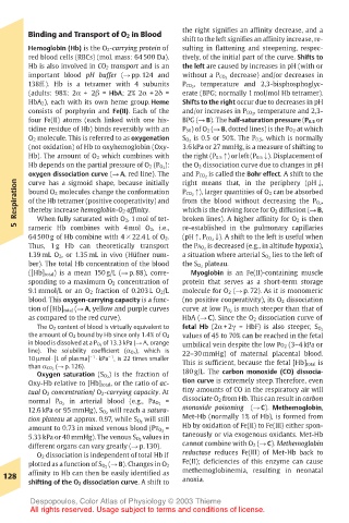
Page 141 - Color_Atlas_of_Physiology_5th_Ed._-_A._Despopoulos_2003
P. 141
Binding and Transport of O 2 in Blood  the right signifies an affinity decrease, and a
                                       shift to the left signifies an affinity increase, re-
       Hemoglobin (Hb) is the O 2-carrying protein of  sulting in flattening and steepening, respec-
       red blood cells (RBCs) (mol. mass: 64 500 Da).  tively, of the initial part of the curve. Shifts to
       Hb is also involved in CO 2 transport and is an  the left are caused by increases in pH (with or
       important blood pH buffer (! pp. 124 and  without a P CO 2 decrease) and/or decreases in
       138ff.). Hb is a tetramer with 4 subunits  P CO 2 , temperature and 2,3-bisphosphoglyc-
       (adults: 98%: 2α + 2" = HbA; 2% 2α + 2δ =  erate (BPG; normally 1 mol/mol Hb tetramer).
       HbA 2), each with its own heme group. Heme  Shifts to the right occur due to decreases in pH
       consists of porphyrin and Fe(II). Each of the  and/or increases in P CO 2 , temperature and 2,3-
       four Fe(II) atoms (each linked with one his-  BPG (! B). The half-saturation pressure (P 0.5 or
       tidine residue of Hb) binds reversibly with an  P 50) of O 2 (! B, dotted lines) is the PO 2 at which
       O 2 molecule. This is referred to as oxygenation  S O 2 is 0.5 or 50%. The P 0.5, which is normally
       (not oxidation) of Hb to oxyhemoglobin (Oxy-  3.6 kPa or 27 mmHg, is a measure of shifting to
       Hb). The amount of O 2 which combines with  the right (P 0.5") or left (P 0.5#). Displacement of
       Hb depends on the partial pressure of O 2 (P O 2 ):  the O 2 dissociation curve due to changes in pH
       oxygen dissociation curve (! A, red line). The  and P CO 2 is called the Bohr effect. A shift to the
    Respiration  of the Hb tetramer (positive cooperativity) and  P CO 2 "), larger quantities of O 2 can be absorbed
                                       right means that, in the periphery (pH#,
       curve has a sigmoid shape, because initially
       bound O 2 molecules change the conformation
                                       from the blood without decreasing the P O 2 ,
                                       which is the driving force for O 2 diffusion (! B,
       thereby increase hemoglobin-O 2 affinity.
         When fully saturated with O 2, 1 mol of tet-
    5  rameric Hb combines with 4 mol O 2, i.e.,  broken lines). A higher affinity for O 2 is then
                                       re-established in the pulmonary capillaries
       64 500 g of Hb combine with 4 ! 22.4 L of O 2.  (pH", P CO 2 #). A shift to the left is useful when
       Thus, 1 g Hb can theoretically transport  the PA O 2 is decreased (e.g., in altitude hypoxia),
       1.39 mL O 2, or 1.35 mL in vivo (Hüfner num-  a situation where arterial S O 2 lies to the left of
       ber). The total Hb concentration of the blood  the S O 2 plateau.
       ([Hb] total) is a mean 150 g/L (! p. 88), corre-  Myoglobin is an Fe(II)-containing muscle
       sponding to a maximum O 2 concentration of  protein that serves as a short-term storage
       9.1 mmol/L or an O 2 fraction of 0.203 L O 2/L  molecule for O 2 (! p. 72). As it is monomeric
       blood. This oxygen-carrying capacity is a func-  (no positive cooperativity), its O 2 dissociation
       tion of [Hb] total (! A, yellow and purple curves  curve at low P O 2 is much steeper than that of
       as compared to the red curve).  HbA (! C). Since the O 2 dissociation curve of
         The O 2 content of blood is virtually equivalent to  fetal Hb (2α + 2γ = HbF) is also steeper, S O 2
                                       values of 45 to 70% can be reached in the fetal
       the amount of O 2 bound by Hb since only 1.4% of O 2
       in blood is dissolved at a P O 2 of 13.3 kPa (! A, orange  umbilical vein despite the low PO 2 (3–4 kPa or
       line). The solubility coefficient (α O 2 ), which is  22–30 mmHg) of maternal placental blood.
       10µmol ! [L of plasma] – 1 ! kPa , is 22 times smaller
                       – 1
       than α CO 2 (! p. 126).         This is sufficient, because the fetal [Hb] total is
         Oxygen saturation (S O 2 ) is the fraction of  180 g/L. The carbon monoxide (CO) dissocia-
       Oxy-Hb relative to [Hb] total, or the ratio of ac-  tion curve is extremely steep. Therefore, even
       tual O 2 concentration/ O 2-carrying capacity. At  tiny amounts of CO in the respiratory air will
       normal P O 2 in arterial blood (e.g., Pa O 2 =  dissociate O 2 from Hb. This can result in carbon
       12.6 kPa or 95 mmHg), S O 2 will reach a satura-  monoxide poisoning  (! C). Methemoglobin,
       tion plateau at approx. 0.97, while S O 2 will still  Met-Hb (normally 1% of Hb), is formed from
       amount to 0.73 in mixed venous blood (PV O 2 =  Hb by oxidation of Fe(II) to Fe(III) either spon-
       5.33 kPa or 40 mmHg). The venous S O 2 values in  taneously or via exogenous oxidants. Met-Hb
       different organs can vary greatly (! p. 130).  cannot combine with O 2 (! C). Methemoglobin
         O 2 dissociation is independent of total Hb if  reductase reduces Fe(III) of Met-Hb back to
                                       Fe(II); deficiencies of this enzyme can cause
       plotted as a function of S O 2 (! B). Changes in O 2  methemoglobinemia, resulting in neonatal
  128  affinity to Hb can then be easily identified as  anoxia.
       shifting of the O 2 dissociation curve. A shift to
       Despopoulos, Color Atlas of Physiology © 2003 Thieme
       All rights reserved. Usage subject to terms and conditions of license.