
Page 258 - Color_Atlas_of_Physiology_5th_Ed._-_A._Despopoulos_2003
P. 258
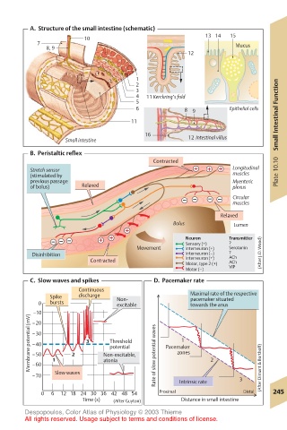
A.  Structure of the small intestine (schematic)
                      10                             13 14  15
          7                                                 Mucus
             8, 9
                                                12

                                   1
                                   2
                                   3
                                   4  11 Kerckring’s fold
                                   5
                                   6           8  9        Epithelial cells
                                  11                                   Small Intestinal Function

                                     16         12 Intestinal villus
                 Small intestine
         B.  Peristaltic reflex
                                       Contracted
         Stretch sensor                                    Longitudinal  Plate 10.10
         (stimulated by                                    muscles
         previous passage                                  Myenteric
         of bolus)    Relaxed                              plexus
                                                           Circular
                                                           muscles
                                                        Relaxed
                                            Bolus           Lumen

                                                Neuron    Transmitter
                                                Sensory (+)  ?
                                   Movement     Interneuron (+)  Serotonin
         Disinhibition                          Interneuron (–)  ? ACh  (After J.D.Wood)
                        Contracted              Interneuron (+)  ACh
                                                Motor, type 2 (+)
                                                Motor (–)  VIP
         C.  Slow waves and spikes      D.  Pacemaker rate
                     Continuous
              Spike  discharge  Non-             Maximal rate of the respective
                                                 pacemaker situated
           0  bursts          excitable          towards the anus
         –10
        Membrane potential (mV)  –30  2  3  Non-excitable,  Rate of slow potential waves  Pacemaker
         –20
                                               1
                             Threshold
         –40
                             potential
                                              zones
         –50
         –60
         –70  1 Slow waves  atonia            Intrinsic rate  2  3  (After Dimant & Borthoff)
           0  6  12 18 24 30 36 42 48 54  Proximal            Distal  245
                      Time (s)  (After Guyton)  Distance in small intestine
       Despopoulos, Color Atlas of Physiology © 2003 Thieme
       All rights reserved. Usage subject to terms and conditions of license.