
Page 358 - Color_Atlas_of_Physiology_5th_Ed._-_A._Despopoulos_2003
P. 358
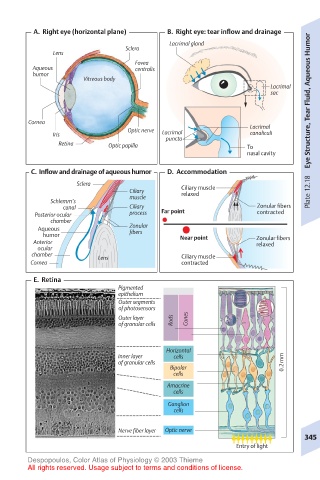
A.  Right eye (horizontal plane)  B.  Right eye: tear inflow and drainage
                                       Lacrimal gland
                             Sclera
             Lens
                               Fovea
        Aqueous                centralis
        humor
                    Vitreous body
                                                              Lacrimal
                                                              sac      Eye Structure, Tear Fluid, Aqueous Humor

       Cornea
                              Optic nerve                Lacrimal
             Iris                     Lacrimal           canaliculi
                                      puncta
              Retina     Optic papilla                   To
                                                         nasal cavity
        C.  Inflow and drainage of aqueous humor  D.  Accommodation
                  Sclera
                              Ciliary     Ciliary muscle               Plate 12.18
                                          relaxed
                              muscle
            Schlemm’s
               canal          Ciliary                      Zonular fibers
         Posterior ocular     process  Far point           contracted
            chamber
          Aqueous             Zonular
           humor              fibers      Near point       Zonular fibers
         Anterior                                          relaxed
         ocular
        chamber        Lens               Ciliary muscle
        Cornea                            contracted
         E.  Retina
                            Pigmented
                            epithelium
                            Outer segments
                            of photosensors
                            Outer layer  Rods  Cones
                            of granular cells


                                      Horizontal
                            Inner layer  cells
                            of granular cells                    0.2mm
                                       Bipolar
                                        cells
                                       Amacrine
                                        cells
                                       Ganglion
                                        cells

                            Nerve fiber layer  Optic nerve
                                                                      345
                                                      Entry of light
       Despopoulos, Color Atlas of Physiology © 2003 Thieme
       All rights reserved. Usage subject to terms and conditions of license.