
Page 357 - Color_Atlas_of_Physiology_5th_Ed._-_A._Despopoulos_2003
P. 357
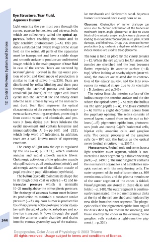
Eye Structure, Tear Fluid,      lar meshwork and Schlemm’s canal. Aqueous
       Aqueous Humor                   humor is renewed once every hour or so.
                                       Glaucoma. Obstruction of humor drainage can
       Light entering the eye must pass through the  occur due to chronic obliteration of the trabecular
       cornea, aqueous humor, lens and vitreous body,  meshwork (open-angle glaucoma) or due to acute
       which are collectively called the optical ap-  block of the anterior angle (angle-closure glaucoma)
       paratus, before reaching the retina and its  leading to elevated intraocular pressure, pain, retinal
                                       damage, and blindness. Drugs that decrease humor
       light-sensitive photosensors (! A). This pro-
    Central Nervous System and Senses  must be transparent and have a stable shape  The lens is held in place by the ciliary zonules
                                       production (e.g. carbonic anhydrase inhibitors) and
       duces a reduced and inverse image of the visual
                                       induce meiosis are used to treat glaucoma.
       field on the retina. All parts of the apparatus
       and smooth surface to produce an undistorted
                                       (! C). When the eye adjusts for far vision, the
       image, which is the main purpose of tear fluid
                                       zonules are stretched and the lens becomes
       in case of the cornea. Tears are secreted by
                                       flatter, especially its anterior surface (! D,
       lacrimal glands located in the top outer por-
                                       top). When looking at nearby objects (near vi-
       tion of orbit and their mode of production is
                                       sion), the zonules are relaxed due to contrac-
       similar to that of saliva (! p. 236). Tears are
                                       tion of the ciliary muscle, and the lens reas-
       distributed by reflex blinking and then pass
                                       sumes its original shape due to its elasticity
       through the lacrimal puncta and lacrimal
                                        The retina lines the interior surface of the
       eyelid into the lacrimal sac and finally drain
                                       bulbus except the anterior surface and the site
       into the nasal sinuses by way of the nasolacri-
                                       where the optical nerve (! A) exits the bulbus
    12  canaliculi (or ducts) of the upper and lower  (! D , bottom, and p. 346).
       mal duct. Tear fluid improves the optical
                                       via the optic papilla (! A). The fovea centralis
       characteristics of the cornea by smoothing un-  (! A) forms a slight depression across from
       even surfaces, washing away dust, protecting it  the pupillary opening. The retina consists of
       from caustic vapors and chemicals, and pro-  several layers, named from inside out as fol-
       tects it from drying out. Tears lubricate the  lows (! E): pigmented epithelium, photosen-
       eyelid movement and contain lysozyme and  sors (rods and cones), Cajal’s horizontal cells,
       immunoglobulin A (! pp. 96ff. and 232),  bipolar cells, amacrine cells, and ganglion
       which help ward off infections. In addition,  cells. The central processes of the ganglion
       tears are a well known mode of expressing  cells (n ! 10 ) exit the bulbus as the optical
                                               6
       emotions.                       nerve (retinal circuitry; ! p. 355ff.).
         The entry of light into the eye is regulated  Photosensors. Retinal rods and cones have a
       by the iris (! A; p. 353 C1), which contains  light-sensitive outer segment, which is con-
       annular and radial smooth muscle fibers.  nected to a inner segment by a thin connecting
       Cholinergic activation of the sphincter muscle  part (! p. 349 C1). The inner segment contains
       of pupil leads to pupil contraction (miosis), and  the normal cell organelles and establishes syn-
       adrenergic activation of the dilator muscle of  aptic contact with the neighboring cells. The
       pupil results in pupil dilatation (mydriasis).  outer segment of the rod cells contains ca. 800
         The bulbus (eyeball) maintains its shape due  membranous disks, and the plasma membrane
       to its tough outer coat or sclera (! C) and in-  of the outer segment of the cones is folded.
       traocular  pressure  which  is  normally  Visual pigments are stored in these disks and
       10–21 mmHg above the atmospheric pressure.  folds (! p. 348). The outer segment is continu-
       The drainage of aqueous humor must balance  ously regenerated; the old membranous disks
       its production to maintain a constant ocular  at the tip of the cell are shed and replaced by
       pressure (! C). Aqueous humor is produced in  new disks from the inner segment. The phago-
       the ciliary process of the posterior ocular cham-  cytic cells of the pigmented epithelium engulf
       ber with the aid of carbonic anhydrase and ac-  the disks shed by the rods in the morning, and
       tive ion transport. It flows through the pupil  those shed by the cones in the evening. Some
       into the anterior ocular chamber and drains  ganglion cells contain a light-sensitive pig-
  344  into the venous system by way of the trabecu-  ment (! p. 334).
       Despopoulos, Color Atlas of Physiology © 2003 Thieme
       All rights reserved. Usage subject to terms and conditions of license.