
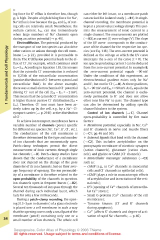
Page 47 - Color_Atlas_of_Physiology_5th_Ed._-_A._Despopoulos_2003
P. 47
!
                +
       ing force for K efflux is therefore low, though  can either be left intact, or a membrane patch
       g K is high. Despite a high driving force for Na , +  can excised for isolated study (! B1). In single-
       Na influx is low because the g Na and f Na of rest-  channel recording, the membrane potential is
         +
       ing cells are relatively small. Nonetheless, the  kept at a preset value (voltage clamp). This per-
       sodium current, I Na, can rise tremendously  mits the measurement of ionic current in a
       when large numbers of Na channels open  single channel. The measurements are plotted
                         +
       during an action potential (! p. 46).  (! B3) as current (I) over voltage (V). The slope
         Electrodiffusion. The potential produced by  of the I/V curve corresponds to the conduct-
                                       ance of the channel for the respective ion spe-
       the transport of one ion species can also drive
    Fundamentals and Cell Physiology  flux of Cl , for example, which continues until  intercepts the x-axis of the curve (I = 0). The +
                                       cies (see Eq. 1.18). The zero-current potential is
       other cations or anions through the cell mem-
       brane (! p. 22), provided it is permeable to
                                       defined as the voltage at which the I/V curve
              +
       them. The K diffusion potential leads to the ef-
             –
                                       ion species producing current I can be deduced
                                       from the zero-current potential. In example B,
       E Cl = E m. According to Equation 1.18, this means
       that the cytosolic Cl concentration is reduced
                   –
                                       the zero-current potential equals – 90 mV.
                                       Under the conditions of this experiment, an
       to 1/25 th of the extracellular concentration
                      –
                                       electrochemical gradient exists only for Na
       (passive distribution of Cl between cytosol and
       extracellular fluid). In the above example,
                                                   –
                                          +
                                       and K , but not for Cl (! B). At these gradients,
                             –
                                       E K = – 90 mV and E Na = + 90 mV. As E K equals the
       there was a small electrochemical Cl potential
             –
                                                     +
                          –
       This means that the cytosolic Cl concentration
                                       sively permeable to K and does not allow
                                                  +
                                       other ions like Na to pass. The channel type
                        –
       is higher than in passive Cl distribution (E Cl =
       E m). Therefore, Cl ions must have been ac-
    1  driving Cl out of the cell (E m – E Cl = – 2 mV).  zero-current potential, the channel is exclu-
                  –
                                       can also be determined by adding specific
       tively taken up by the cell, e.g., by a Na - Cl –  channel blockers to the system.
                                 +
       symport carrier (! p. 29 B): active distribution  Control of ion channels (! C). Channel
       of Cl .                         open-probability is controlled by five main
          –
         To achieve ion transport, membranes have a  factors:
       variable number of channels (pores) specific  ! Membrane potential, especially in Na , Ca 2+
                                                                +
                           2+
                                –
                             +
                        +
                                           +
       for different ion species (Na , Ca , K , Cl , etc.).  and K channels in nerve and muscle fibers
       The conductance of the cell membrane is  (! C1; pp. 46 and 50).
       therefore determined by the type and number  ! External ligands that bind with the channel
       of ion channels that are momentarily open.  (! C2). This includes acetylcholine on the
       Patch–clamp techniques permit the direct  postsynaptic membrane of nicotinic synapses
       measurement of ionic currents through single  (cation channels), glutamate (cation chan-
                                                         –
       ion channels (! B). Patch–clamp studies have  nels), and glycine or GABA (Cl channels).
       shown that the conductance of a membrane  ! Intracellular messenger substances (! C3)
       does not depend on the change of the pore  such as:
       diameter of its ion channels, but on their aver-  — cAMP (e.g., in Ca 2+  channels in myocardial
                                               –
       age frequency of opening. The ion permeabil-  cells and Cl channels in epithelial cells);
       ity of a membrane is therefore related to the  — cGMP (plays a role in muscarinergic effects
       open-probability of the channels in question.  of acetylcholine and in excitation of the reti-
       Ion channels open in frequent bursts (! B2).  nal rods);
       Several ten thousands of ions pass through the  — IP3 (opening of Ca 2+  channels of intracellu-
       channel during each individual burst, which  lar Ca 2+ stores);
       lasts for only a few milliseconds.  — Small G-proteins (Ca 2+  channels of the cell
         During a patch–clamp recording, the open-  membrane);
       ing (0.3–3 µm in diameter) of a glass electrode  — Tyrosine kinases (Cl –  and K +  channels
       is placed over a cell membrane in such a way  during apoptosis);
                                                 +
       that the opening covers only a small part of the  — Ca 2+ (affects K channels and degree of acti-
                                                    +
       membrane (patch) containing only one or a  vation of rapid Na channels; ! p. 46).
   34  small number of ion channels. The whole cell
       Despopoulos, Color Atlas of Physiology © 2003 Thieme
       All rights reserved. Usage subject to terms and conditions of license.