
Page 265 - Hall et al (2015) Principles of Critical Care-McGraw-Hill
P. 265
CHAPTER 25: Cardiopulmonary Resuscitation 169
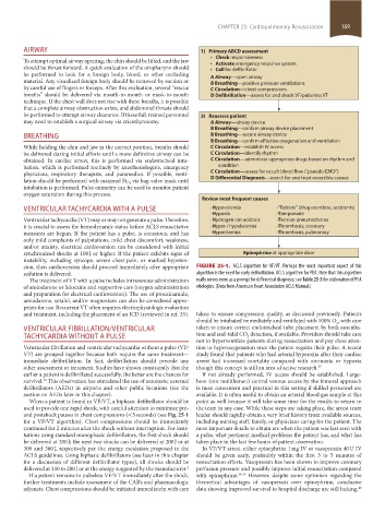
AIRWAY 1) Primary ABCD assessment
Check responsiveness
To attempt optimal airway opening, the chin should be lifted, and the jaw
should be thrust forward. A quick evaluation of the oropharynx should Activate emergency response system
Call for defibrillator
be performed to look for a foreign body, blood, or other occluding
material. Any visualized foreign body should be removed by suction or A Airway—open airway
B Breathing—positive pressure ventilations
by careful use of fingers or forceps. After this evaluation, several “rescue C Circulation—chest compressions
breaths” should be delivered via mouth-to-mouth or mask-to-mouth D Defibrillation—assess for and shock VF/pulseless VT
technique. If the chest wall does not rise with these breaths, it is possible
that a complete airway obstruction exists, and abdominal thrusts should
be performed to attempt airway clearance. If these fail, trained personnel 2) Reassess patient
may need to establish a surgical airway via cricothyrotomy. A Airway—airway device
B Breathing—confirm airway device placement
BREATHING B Breathing—secure airway device
B Breathing—confirm e ective oxygenation and ventilation
While holding the chin and jaw in the correct position, breaths should C Circulation—establish IV access
be delivered during initial efforts until a more definitive airway can be C Circulation—identify rhythm
obtained. In cardiac arrest, this is performed via endotracheal intu- C Circulation—administer appropriate drugs based on rhythm and
bation, which is performed routinely by anesthesiologists, emergency condition
physicians, respiratory therapists, and paramedics. If possible, venti- C Circulation—assess for occult blood flow (“pseudo-EMD”)
via bag-valve mask until D Di erential Diagnosis—search for and treat reversible causes
lation should be performed with maximal Fi O 2
intubation is performed. Pulse oximetry can be used to monitor patient
oxygen saturation during this process.
Review most frequent causes
VENTRICULAR TACHYCARDIA WITH A PULSE -Hypovolemia -“Tablets” (drug overdose, accidents)
-Hypoxia -Tamponade
Ventricular tachycardia (VT) may or may not generate a pulse. Therefore, -Hydrogen ion-acidosis -Tension pneumothorax
it is crucial to assess the hemodynamic status before ACLS resuscitative -Hyper-/Hypokalemia -Thrombosis, coronary
measures are begun. If the patient has a pulse, is conscious, and has -Hypothermia -Thrombosis, pulmonary
only mild complaints of palpitations, mild chest discomfort, weakness,
and/or anxiety, electrical cardioversion can be considered with initial
synchronized shocks at 100 J or higher. If the patient exhibits signs of Epinephrine at appropriate dose
instability, including syncope, severe chest pain, or marked hypoten-
sion, then cardioversion should proceed immediately after appropriate FIGURE 25-1. ACLS algorithm for VF/VT. Perhaps the most important aspect of this
sedation is delivered. algorithm is the need for early defibrillation. ACLS algorithm for PEA. Note that this algorithm
The treatment of VT with a pulse includes intravenous administration really serves more as a prompt for differential diagnosis; see Table 25-3 for elaboration of PEA
of amiodarone or lidocaine and supportive care (oxygen administration etiologies. (Data from American Heart Association ACLS Manual.)
and preparation for electrical cardioversion). The use of procainamide,
amiodarone, sotalol, and/or magnesium can also be considered appro-
priate for use. Recurrent VT often requires electrophysiologic evaluation
and treatment, including the placement of an ICD (reviewed in ref. 35). taken to ensure compression quality, as discussed previously. Patients
should be intubated immediately and ventilated with 100% O , with care
2
VENTRICULAR FIBRILLATION/VENTRICULAR taken to ensure correct endotracheal tube placement by both ausculta-
TACHYCARDIA WITHOUT A PULSE tion and end-tidal CO detection, if available. Providers should take care
2
not to hyperventilate patients during resuscitation and pay close atten-
Ventricular fibrillation and ventricular tachycardia without a pulse (VF/ tion to hyperoxygenation once the patient regains their pulse. A recent
VT) are grouped together because both require the same treatment— study found that patients who had arterial hyperoxia after their cardiac
immediate defibrillation. In fact, defibrillation should precede any arrest had increased mortality compared with normoxia or hypoxia
other assessment or treatment. Studies have shown consistently that the though this concept is still an area of active research. 37
earlier a patient is defibrillated successfully, the better are the chances for If not already performed, IV access should be established. Large-
survival. This observation has stimulated the use of automatic external bore (not multilumen) central venous access by the femoral approach
36
defibrillators (AEDs) in airports and other public locations (see the is most convenient and practical in this setting if skilled personnel are
section on AEDs later in this chapter). available. It is often useful to obtain an arterial blood gas sample at this
When a patient is found in VF/VT, a biphasic defibrillator should be point as well because it will take some time for the results to return to
used to provide one rapid shock, with careful attention to minimize pre- the team in any case. While these steps are taking place, the arrest team
and postshock pauses in chest compressions (<5 seconds) (see Fig. 25-1 leader should rapidly obtain a very brief history from available sources,
for a VF/VT algorithm). Chest compressions should be immediately including nursing staff, family, or physicians caring for the patient. The
continued for 2 minutes after the shock without interruption. For insti- most important details to obtain are when the patient was last seen with
tutions using standard monophasic defibrillators, the first shock should a pulse, what pertinent medical problems the patient has, and what has
be delivered at 200 J; the next two shocks can be delivered at 200 J or at taken place in the last few hours of patient observation.
300 and 360 J, respectively per the energy escalation proposed in the In VF/VT arrest, either epinephrine 1 mg IV or vasopressin 40 U IV
ACLS guidelines. Using biphasic defibrillators (see later in this chapter should be given early, preferably within the first 3 to 5 minutes of
for a discussion of different defibrillator types), all shocks should be resuscitation efforts. Vasopressin has been shown to improve coronary
delivered at 150 to 200 J or at the energy suggested by the manufacturer. 4 perfusion pressure and possibly improve initial resuscitation compared
If a patient remains in pulseless VF/VT immediately after the shock, with epinephrine. 38-39 However, despite some optimism regarding the
further treatments include assessment of the CAB’s and pharmacologic theoretical advantages of vasopressin over epinephrine, conclusive
adjuncts. Chest compressions should be initiated immediately, with care data showing improved survival to hospital discharge are still lacking.
40
section02.indd 169 1/13/2015 2:05:10 PM