
Page 267 - Hall et al (2015) Principles of Critical Care-McGraw-Hill
P. 267
CHAPTER 25: Cardiopulmonary Resuscitation 171
1) Definition 1) Primary ABCD assessment
Slow: absolute bradycardia = <60 bpm Check responsiveness
or Activate emergency response system
Relatively slow: rate less than expected relative to condition or Call for defibrillator
cause
A Airway—open airway
B Breathing—positive pressure ventilations
2) Primary ABCD assessment C Circulation—chest compressions
Assess ABCs C Confirm true asystole
Secure airway noninvasively D Defibrillation—assess for and shock VF/pulseless VT
Ensure availability of monitor/defibrillator
2) Check rhythm
3) Reassess patient 3) Persistent or recurrent VF/VT
Assess secondary ABCs (need invasive airway?)
Oxygen—IV access—monitor—fluids 4) Re-assess patient
Vital signs, pulse oximeter, monitor blood pressure
Obtain and review 12-lead ECG A Airway—airway device
Obtain and review portable chest x-ray B Breathing—confirm airway device placement
Problem-focused history B Breathing—secure airway device
Problem-focused physical examination B Breathing—confirm effective oxygenation and ventilation
Consider causes (di erential diagnosis) C Circulation—establish IV access
C Circulation—identify rhythm
C Circulation—administer appropriate drugs based on rhythm
4) Serious signs or symptoms due to the bradycardia? and condition
D Differential diagnosis—search for and treat reversible causes
6) Type II second-degree AV block 5) Intervention sequence 5) Epinephrine or vasopressin at appropriate dose
or Epinephrine
Third-degree AV block? Transcutaneous pacing 6) Resume attempts to defibrillate
Dopamine 7) Consideration of antiarrythmics
Isoproterenol
Amiodarone
Lidocaine
Observe Prepare for transvenous pacing Magnesium
If symptoms develop, use transcutaneous Procainamide
pacemaker until pacer placed
8) Resume attempts to defibrillate
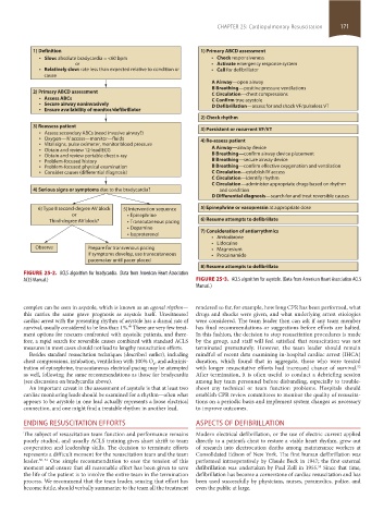
FIGURE 25-2. ACLS algorithm for bradycardia. (Data from American Heart Association
ACLS Manual.) FIGURE 25-3. ACLS algorithm for asystole. (Data from American Heart Association ACLS
Manual.)
complex can be seen in asystole, which is known as an agonal rhythm— rendered so far, for example, how long CPR has been performed, what
this carries the same grave prognosis as asystole itself. Unwitnessed drugs and shocks were given, and what underlying arrest etiologies
cardiac arrest with the presenting rhythm of asystole has a dismal rate of were considered. The team leader then can ask if any team member
survival, usually considered to be less than 1%. There are very few treat- has final recommendations or suggestions before efforts are halted.
49
ment options for rescuers confronted with asystolic patients, and there- In this fashion, the decision to stop resuscitation procedures is made
fore, a rapid search for reversible causes combined with standard ACLS by the group, and staff will feel satisfied that resuscitation was not
measures in most cases should not lead to lengthy resuscitation efforts. terminated prematurely. However, the team leader should remain
Besides standard resuscitation techniques (described earlier), including mindful of recent data examining in-hospital cardiac arrest (IHCA)
chest compressions, intubation, ventilation with 100% O , and adminis- duration, which found that in aggregate, those who were treated
2
tration of epinephrine, transcutaneous electrical pacing may be attempted with longer resuscitative efforts had increased chance of survival.
52
as well, following the same recommendations as those for bradycardia After termination, it is often useful to conduct a debriefing session
(see discussion on bradycardia above). among key team personnel before disbanding, especially to trouble-
An important caveat in the assessment of asystole is that at least two shoot any technical or team function problems. Hospitals should
cardiac monitoring leads should be examined for a rhythm—often what establish CPR review committees to monitor the quality of resuscita-
appears to be asystole in one lead actually represents a loose electrical tions on a periodic basis and implement system changes as necessary
connection, and one might find a treatable rhythm in another lead. to improve outcomes.
ENDING RESUSCITATION EFFORTS ASPECTS OF DEFIBRILLATION
The subject of resuscitation team function and performance remains Modern electrical defibrillation, or the use of electric current applied
poorly studied, and usually ACLS training gives short shrift to team directly to a patient’s chest to restore a viable heart rhythm, grew out
cooperation and leadership skills. The decision to terminate efforts of research into electrocution deaths among maintenance workers at
represents a difficult moment for the resuscitation team and the team Consolidated Edison of New York. The first human defibrillation was
leader. 50, 51 One simple recommendation to ease the tension of this performed intraoperatively by Claude Beck in 1947; the first external
moment and ensure that all reasonable effort has been given to save defibrillation was undertaken by Paul Zoll in 1955. Since that time,
53
the life of the patient is to involve the entire team in the termination defibrillation has become a cornerstone of cardiac resuscitation and has
process. We recommend that the team leader, sensing that effort has been used successfully by physicians, nurses, paramedics, police, and
become futile, should verbally summarize to the team all the treatment even the public at large.
section02.indd 171 1/13/2015 2:05:12 PM