
Page 266 - Hall et al (2015) Principles of Critical Care-McGraw-Hill
P. 266
170 PART 2: General Management of the Patient
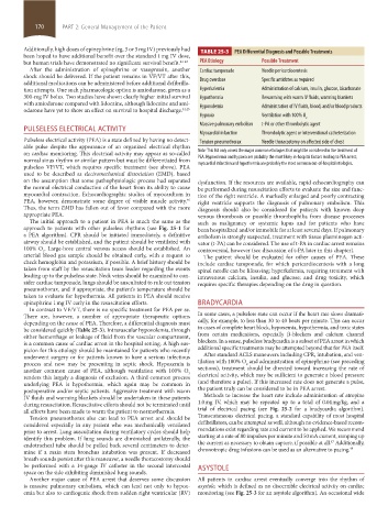
Additionally, high doses of epinephrine (eg, 3 or 5 mg IV) previously had TABLE 25-3 PEA Differential Diagnosis and Possible Treatments
been hoped to have additional benefit over the standard 1 mg IV dose,
but human trials have demonstrated no significant survival benefit. 41-43 PEA Etiology Possible Treatment
After the administration of epinephrine or vasopressin, another Cardiac tamponade Needle pericardiocentesis
shock should be delivered. If the patient remains in VF/VT after this,
additional medications can be administered before additional defibrilla- Drug overdose Specific antidotes as required
tion attempts. One such pharmacologic option is amiodarone, given as a Hyperkalemia Administration of calcium, insulin, glucose, bicarbonate
300-mg IV bolus. Two studies have shown clearly higher initial survival Hypothermia Rewarming with warm IV fluids, warming blankets
with amiodarone compared with lidocaine, although lidocaine and ami-
odarone have yet to show an effect on survival to hospital discharge. 44,45 Hypovolemia Administration of IV fluids, blood, and/or blood products
Hypoxia Ventilation with 100% O
2
Massive pulmonary embolism t-PA or other thrombolytic agent
PULSELESS ELECTRICAL ACTIVITY
Myocardial infarction Thrombolytic agent or interventional catheterization
Pulseless electrical activity (PEA) is a state defined by having no detect- Tension pneumothorax Needle thoracostomy on affected side of chest
able pulse despite the appearance of an organized electrical rhythm
on cardiac monitoring. This electrical activity may appear as so-called Note: This list only covers the major common etiologies that might be considered in the treatment of
normal sinus rhythm or similar pattern but must be differentiated from PEA. Hypovolemia and hypoxia are probably the most likely in-hospital factors leading to PEA arrest;
pulseless VF/VT, which requires specific treatment (see above). PEA myocardial infarction and hypothermia are probably the most common out-of-hospital etiologies.
used to be described as electromechanical dissociation (EMD), based
on the assumption that some pathophysiologic process had separated dysfunction. If the resources are available, rapid echocardiography can
the normal electrical conduction of the heart from its ability to cause be performed during resuscitation efforts to evaluate the size and func-
myocardial contraction. Echocardiographic studies of myocardium in tion of the right ventricle. A markedly enlarged and poorly contracting
PEA, however, demonstrate some degree of visible muscle activity. right ventricle supports the diagnosis of pulmonary embolism. This
46
Thus, the term EMD has fallen out of favor compared with the more diagnosis should also be considered for patients with known deep
appropriate PEA. venous thrombosis or possible thrombophilia from disease processes
The initial approach to a patient in PEA is much the same as the such as malignancy or systemic lupus and for patients who have
approach to patients with other pulseless rhythms (see Fig. 25-1 for been hospitalized and/or immobile for at least several days. If pulmonary
a PEA algorithm). CPR should be initiated immediately, a definitive embolism is strongly suspected, treatment with tissue plasminogen acti-
airway should be established, and the patient should be ventilated with vator (t-PA) can be considered. The use of t-PA in cardiac arrest remains
100% O . Large-bore central venous access should be established. An controversial, however (see discussion of t-PA later in this chapter).
2
arterial blood gas sample should be obtained early, with a request to The patient should be evaluated for other causes of PEA. These
check hemoglobin and potassium, if possible. A brief history should be include cardiac tamponade, for which pericardiocentesis with a long
taken from staff by the resuscitation team leader regarding the events spinal needle can be lifesaving; hyperkalemia, requiring treatment with
leading up to the pulseless state. Neck veins should be examined to con- intravenous calcium, insulin, and glucose; and drug toxicity, which
sider cardiac tamponade, lungs should be auscultated to rule out tension requires specific therapies depending on the drug in question.
pneumothorax, and if appropriate, the patient’s temperature should be
taken to evaluate for hypothermia. All patients in PEA should receive
epinephrine 1 mg IV early in the resuscitation efforts. BRADYCARDIA
In contrast to VF/VT, there is no specific treatment for PEA per se.
There are, however, a number of appropriate therapeutic options In some cases, a pulseless state can occur if the heart rate slows dramati-
depending on the cause of PEA. Therefore, a differential diagnosis must cally, for example, to less than 30 to 40 beats per minute. This can occur
be considered quickly (Table 25-3). Intravascular hypovolemia, through in cases of complete heart block, hypoxemia, hypothermia, and toxic states
either hemorrhage or leakage of fluid from the vascular compartment, from certain medications, especially β-blockers and calcium channel
is a common cause of cardiac arrest in the hospital setting. A high sus- blockers. In a sense, pulseless bradycardia is a subset of PEA arrest in which
picion for this etiology should be maintained for patients who recently additional specific treatments may be attempted beyond that for PEA itself.
underwent surgery or for patients known to have a serious infectious After standard ACLS maneuvers including CPR, intubation, and ven-
process and now may be presenting in septic shock. Hypoxemia is tilation with 100% O and administration of epinephrine (see preceding
2
another common cause of PEA, although ventilation with 100% O sections), treatment should be directed toward increasing the rate of
2
renders this largely a diagnosis of exclusion. A third common process electrical activity, which may be sufficient to generate a blood pressure
underlying PEA is hypothermia, which again may be common in (and therefore a pulse). If this increased rate does not generate a pulse,
postoperative and/or septic patients. Aggressive treatment with warm the patient truly can be considered to be in PEA arrest.
IV fluids and warming blankets should be undertaken in these patients Methods to increase the heart rate include administration of atropine
during resuscitation. Resuscitative efforts should not be terminated until 1.0 mg IV, which may be repeated up to a total of 0.04 mg/kg, and a
all efforts have been made to warm the patient to normothermia. trial of electrical pacing (see Fig. 25-2 for a bradycardia algorithm).
Tension pneumothorax also can lead to PEA arrest and should be Transcutaneous electrical pacing, a standard capability of most hospital
considered especially in any patient who was mechanically ventilated defibrillators, can be attempted as well, although no evidence-based recom-
prior to arrest. Lung auscultation during ventilatory cycles should help mendations exist regarding rate and current to be applied. We recommend
identify this problem. If lung sounds are diminished unilaterally, the starting at a rate of 80 impulses per minute and 50 mA current, ramping up
47
endotracheal tube should be pulled back several centimeters to deter- the current as necessary to obtain capture, if possible at all. Additionally,
mine if a main stem bronchus intubation was present. If decreased chronotropic drug infusions can be used as an alternative to pacing. 48
breath sounds persist after this maneuver, a needle thoracostomy should
be performed with a 14-gauge IV catheter in the second intercostal ASYSTOLE
space on the side exhibiting diminished lung sounds.
Another major cause of PEA arrest that deserves some discussion All patients in cardiac arrest eventually converge into the rhythm of
is massive pulmonary embolism, which can lead not only to hypox- asystole, which is defined as no discernible electrical activity on cardiac
emia but also to cardiogenic shock from sudden right ventricular (RV) monitoring (see Fig. 25-3 for an asystole algorithm). An occasional wide
section02.indd 170 1/13/2015 2:05:11 PM