
Page 424 - Hall et al (2015) Principles of Critical Care-McGraw-Hill
P. 424
294 PART 3: Cardiovascular Disorders
Classically, myocardial ischemia has been divided into categories of oxygen supply, particularly in the setting of hemodynamic instability.
including stable angina, unstable angina, and myocardial infarction. These factors include hypotension, decreasing coronary perfusion pres-
Typical angina is exertional, and is relieved promptly by rest or nitro- sure, and tachycardia, limiting diastolic filling time. In addition, anemia
glycerin. Stable angina occurs reproducibly with a similar level of and hypoxemia can limit the amount of oxygen delivered to the heart.
exertion, in a pattern that has not changed over the past 6 months. Coronary vasospasm may also play a role in some patients. Elevation of
Acute coronary syndromes comprise unstable angina and myocardial left ventricular pressures by heart failure can both increase demand and
infarction. Unstable angina consists of ischemic symptoms which are reduce coronary perfusion pressure.
more frequent, severe, or prolonged than the patient’s usual angina, Thus, critically ill patients, usually those with at least some component
are more difficult to control with drugs, or are occurring at rest or with of obstructive coronary artery disease, may develop myocardial isch-
minimal exertion. Cardiac biomarkers are not elevated. Myocardial emia on a hemodynamic basis, with variable contributions of increased
infarction has been classified as “transmural” and “nontransmural,” but demand and decreased supply. On the other hand, catecholamine surges,
this division has been largely abandoned due to the recognition that hemodynamic changes, and inflammatory processes may predispose to
electrocardiographic criteria are neither sensitive nor specific to make rupture of preexisting atherosclerotic plaques. Making the distinction is
this distinction. vital because the treatment is completely different. In the former case,
Acute coronary syndromes were previously classified into Q-wave treatment is aimed at decreasing the oxygen requirement of the myo-
myocardial infarction, non-Q-wave myocardial infarction, and unstable cardium by eliminating provocative stimuli and controlling heart rate
angina. More recently, classification has shifted and has become based and blood pressure, and on optimizing oxygenation and hemoglobin
on the initial electrocardiogram: patients are divided into three groups: concentration. Relief of myocardial ischemia by these measures usually
those with ST elevation (STEMI), without ST elevation but with enzyme results in prompt restoration of left ventricular function without sig-
evidence of myocardial damage (non-ST elevation MI, or non-ST eleva- nificant cellular damage, since the obstruction to flow is ordinarily fixed
tion myocardial infarction [NSTEMI]), and those with unstable angina. and not total. If plaque rupture is playing a role, then simply removing
Classification according to presenting electrocardiogram coincides with or lessening stimuli that increase myocardial oxygen requirements may
current treatment strategies, since patients presenting with ST eleva- not be sufficient to increase the myocardial oxygen supply:demand ratio,
tion benefit from immediate reperfusion and should be treated with and unless attempts are made to reestablish coronary blood flow, signifi-
fibrinolytic therapy or urgent revascularization, whereas fibrinolytic cant myocardial damage may ensue. Antithrombotic and anticoagulant
agents are not effective in other patients with acute coronary syndromes. strategies should be instituted, and consideration of coronary revascu-
The discussion of myocardial infarction in this chapter follows this larization may be indicated.
schematization.
■ PATHOPHYSIOLOGY RECOGNITION OF MYOCARDIAL ISCHEMIA
and demand. The myocardial requirement for oxygen, and hence for ■ SIGNS AND SYMPTOMS
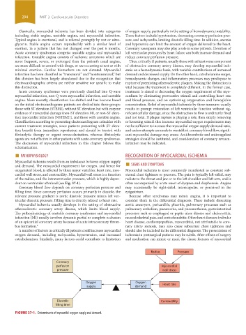
Myocardial ischemia results from an imbalance between oxygen supply
oxygenated blood, is affected by three major variables: heart rate, myo- Myocardial ischemia is most commonly manifested as constant sub-
cardial wall stress, and contractility. Myocardial wall stress is a function sternal chest tightness or pressure. The pain is typically left-sided, may
of the radius, and the intraventricular pressure, which is highly depen- radiate to the throat and jaw or to the left shoulder and left arm, and is
dent on ventricular afterload (see Fig. 37-1). often accompanied by acute onset of dyspnea and diaphoresis. Angina
Coronary blood flow depends on coronary perfusion pressure and may occasionally be right-sided, interscapular, or perceived in the
filling time. Since coronary perfusion occurs primarily in diastole, the epigastrium.
relevant pressure gradient is aortic diastolic pressure minus left ven- Because other syndromes may mimic angina, it is important to
tricular diastolic pressure. Filling time is directly related to heart rate. consider them in the differential diagnosis. These include dissecting
Myocardial ischemia usually develops in the setting of obstructive aortic aneurysm, pericarditis, pleuritis, pulmonary processes such as
atherosclerotic coronary artery disease, which limits blood supply. pulmonary embolism, pneumonia, and pneumothorax, gastrointestinal
The pathophysiology of unstable coronary syndromes and myocardial processes such as esophageal or peptic ulcer disease and cholecystitis,
infarction (MI) usually involves dynamic partial or complete occlusion musculoskeletal pain, and costochondritis. Other heart diseases (valvular
of an epicardial coronary artery because of acute intracoronary throm- heart disease, cardiomyopathies, myocarditis), not attributable to coro-
bus formation. 2 nary artery stenosis, may also cause substernal chest tightness and
A number of factors in critically ill patients could increase myocardial should also be included in the differential diagnosis. The presentation of
oxygen demand, including tachycardia, hypertension, and increased ischemia in postsurgical patients may be subtle. After-effects of surgery
catecholamines. Similarly, many factors could contribute to limitation and medication can mimic or mask the classic features of myocardial
Radius Pressure
Coronary
perfusion Myocardial
pressure wall stress
Oxygen Oxygen Heart rate
supply demand
Diastolic Contractility
filling time
FIGURE 37-1. Determinants of myocardial oxygen supply and demand.
section03.indd 294 1/23/2015 2:07:18 PM