
Page 422 - Hall et al (2015) Principles of Critical Care-McGraw-Hill
P. 422
292 PART 3: Cardiovascular Disorders
V T Onset
v v v v v v v T T T T T T T T T T T
s 8 s 8 s 8 s 8 s 6 s 6 s 4 s 4 s 4 s 4 s 4 s 4 s 4 s 4 s 4 s 4 s 4 s 4
6 7 7 5 0 3 4 2 3 4 4 4 5 5 7 7 8 7
0 0 0 0 0 0 0 0 0 0 0 0 0 0 0 0 0 0
ATP Terminates V T Sinus Rhythm
T 4 T 4 T 4 T 3 T 3 T 3 T 3 T 3 T 1500 v 9 v s 9 v s 9 v s 8 v s 9 v s 8 v s
S 7 D 1 P 0 P 9 P 8 P 7 P 6 P 5 P P 7 1 0 9 1 5
0 0 0 0 0 0 0 0 0 0 0 0 0 0
V T
V T Rx 1 Ramp
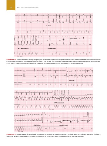
FIGURE 36-10. Example of ventricular antitachycardia pacing (ATP) for ventricular tachycardia (V T). The upper trace is an intracardiac ventricular electrogram (near field); the middle trace
is the electrogram signal recorded from the intracardiac and lead and the ICD can (far field), and the lower trace indicates the marker channel annotations (how the device classifies each beat)
and the interval in milliseconds between successive beats (in milliseconds). TP, antitachycardia pacing; TS, tachycardia sensed; VS, ventricular sensed event.
EGM1: Vtip to Vring
(1 mV)
EGM2: Can to HVB
(1 mV)
V T Onset
Marker Annotation
V–V interval (ms) 1200 v s 1200 v s 1200 v s 7 3 v s 1500 v P 9 0 v s 4 9 v s 7 3 v s 3 v s 3 v s 3 v s 3 v s 3 v s
7
5
7
8
8
0 0 0 0 0 0 0 0 0
ATP Accelerates V T
3 8 s T 3 7 T 3 8 T s 3 8 T 3 T s 3 8 T 3 T s 3 8 T 3 T s 3 8 T s 3 8 T 3 T s 3 8 T 3 T 3 T 3 T 3 T 3 P 4 T 3 T 3 T 3 T P 4 8 v 3 T 2 F 2 F 2 F 3 T 2 F 2 F 3 F 3 F 2 s 9
F 2
T 3
s
P 4
s 9
s 2
s 8
P 4
P 4
s 6
s 8
s 7
P 4
P 4
s 9
s 8
s 8
s 8
s 0
s 1
s 8
D 4
P 4
s 8
s 8
0 0 0 0 0 0 0 0 0 0 0 0 0 0 0 0 0 0 0 0 0 0 0 0 0 0 0 0 0 0 0 0 0 0
V T
V T Rx 1 Burst
Defibrillation Paced Rhythm
F F F v v v v v v v v v c v c v v v v v
s 2 s 2 D 2 s 3 s 2 s 2 s 3 s 2 s 2 s 2 s 2 s 2 E 2 s 3 D 1200 P 1500 P 1500 P 1030 s 1500 P 1350
8 8 6 0 6 8 0 9 9 9 8 5 R 8 0
0 0 VF 0 0 0 0 0 0 0 0 0 0 0 0 20.1 j
VF Rx 1 De b
FIGURE 36-11. Example of ventricular antitachycardia pacing therapy that accelerates the ventricular tachycardia. A 20-J shock converts the arrhythmia to sinus rhythm. The format is
similar to Fig. 36-10. CD, change delivered; FS, ventricular fibrillation sensed; TP, antitachycardia pacing; TS, tachycardia sensed; VS, ventricular sensed event.
section03.indd 292 1/23/2015 2:07:17 PM