
Page 426 - Hall et al (2015) Principles of Critical Care-McGraw-Hill
P. 426
296 PART 3: Cardiovascular Disorders
event, and so critical therapeutic interventions should not be delayed muscle rupture and acute mitral regurgitation, acute ventricular septal
pending assay results. Once elevated, troponin levels can remain high defect, and free wall rupture and tamponade. In some cases, echo-
14
for days to weeks, limiting their utility to detect late reinfarction. cardiography may reveal findings compatible with right ventricular
One challenge with use of troponins in the intensive care unit is that infarction. Echocardiography can also reveal alternative diagnoses,
their elevation may not be confined to acute coronary syndromes. A such as valvular abnormalities, pericardial tamponade, or hypertrophic
number of other conditions prevalent in the critical care setting, includ- cardiomyopathy. Acute right heart failure, manifested by a dilated and
ing sepsis, burns, pulmonary embolism, myocarditis, and renal failure, hypokinetic right ventricle without hypertrophy suggestive of chronic
have been associated with increases in troponin, albeit at levels lower pulmonary hypertension, can suggest pulmonary embolism. 15
than those usually seen with large myocardial infarctions. Detectable Transthoracic echocardiographic images may be suboptimal due to
9
troponin levels in critically ill patients, although they usually emanate a poor acoustic window in critically ill patients, particularly those who
from myocardial cells, may not always represent either irreversible are obese, have chronic lung disease, or are on positive pressure ventila-
cell death or myocardial ischemia. Endotoxin, cytokines, and other tion. Contrast echocardiography may be used to improve image quality.
16
inflammatory mediators, along with catecholamines and conditions Transesophageal echocardiography (TEE) can also provide better visual-
such as hypotension, inotropes or hypoxia may cause the breakdown ization, particularly of valvular structures, and can be performed safely
of cytoplasmic troponin into smaller fragments that can pass through at the bedside.
endothelial monolayers and subsequently be detected by sensitive assays ■
for troponin. In any event, isolated troponin elevation in the absence of HEMODYNAMIC MONITORING
10
ECG changes or other clinical signs of ischemia should be evaluated in In patients with hemodynamic instability that does not improve relatively
the clinical context. In some settings, echocardiography to evaluate for quickly with simple therapeutic maneuvers, invasive hemodynamic
new wall motion abnormalities may be useful. monitoring should be considered. Pulmonary artery catheterization
■ ECHOCARDIOGRAPHY (PAC) provides simultaneous assessment of filling pressures and cardiac
output, and can be quite useful for differential diagnosis in critically ill
To the physician confronted with a critically ill patient, echocar- patients. In patients with hypoxemia and pulmonary infiltrates on chest
diography can be a key element in successful differential diagnosis. x-ray, a frequent dilemma in ICU patients, PAC may be used to differen-
11
Echocardiography is simple, safe, and permits systemic interrogation tiate cardiac from pulmonary causes. Right heart catheterization is also
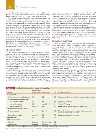
of cardiac chamber size, left and right ventricular function, valvular quite useful in the differential diagnosis of shock. Hemodynamic profiles
structure and motion, atrial size, and the anatomy of the pericardial of patients with different forms of shock are shown in Table 37-1. It is
space. The presence of segmental left ventricular wall motion abnor- important to recognize the possibility of mixed forms of shock in criti-
malities suggests compromise of blood flow to those segments. Doppler cally ill patient. For example, patients with myocardial infarction, even
12
interrogation can be used for noninvasive assessment of right and left in the presence of significant left ventricular dysfunction and suspected
ventricular filling pressures, pulmonary artery pressures, stroke volume, cardiogenic shock, can be relatively volume depleted, perhaps due to
and cardiac output. diaphoresis and/or vomiting. 13,17
Echocardiography is particularly useful in the evaluation of patients Hemodynamic monitoring can also be useful in the diagnosis of
with acute heart failure or suspected cardiogenic shock, and early mechanical complications of infarction, although most causes are more
echocardiography should be routine. Expeditious evaluation of global easily identified with echocardiography. Right heart catheterization may
13
and regional left ventricular performance is crucial for management of reveal a step-up in hemoglobin oxygen saturation diagnostic of ven-
congestive heart failure, with or without suspected myocardial ischemia. tricular septal rupture. The waveform of the PAOP tracing may reveal
Echocardiography is also extremely valuable for the rapid diagnosis of a prominent V wave (10 mm Hg above the mean PAOP is regarded as
mechanical causes of shock after myocardial infarction such as papillary significant) suggesting severe mitral regurgitation, although V waves
TABLE 37-1 Use of Right Heart Catheterization to Diagnose the Etiology of Shock
Pulmonary Artery
Diagnosis Occlusion Pressure Cardiac Output SVR Miscellaneous Comments
Cardiogenic Shock
Cardiogenic shock due to myocardial dysfunction ⇑⇑ ⇓⇓ ⇑⇑ Usually extensive infarction (>40% of LV), severe cardiomyopathy, or myocarditis
Cardiogenic shock due to mechanical defects
Acute ventricular septal defect ⇑ ⇓⇓ ⇑⇑ Oxygen “step-up” at RV level
Acute mitral regurgitation ⇑⇑ Forward CO ⇓⇓ ⇑ V waves in PAOP tracing
Right ventricular infarction Normal or ⇓ ⇓⇓ ⇑⇑ Elevated RA and RV filling pressures with low or normal PAOP
Extracardiac Obstructive Forms of Shock
Pericardial tamponade ⇑ ⇓ or ⇓⇓ ⇑⇑ RA mean, RV end-diastolic pulmonary capillary wedge mean pressures are
elevated and within 5 mm Hg of one another
Massive pulmonary embolism normal or ⇓ ⇓⇓ ⇑⇑ Usual finding is elevated right-sided pressures
Hypovolemic Shock ⇓⇓ ⇓⇓ ⇑⇑
Distributive Forms of Shock
Septic shock ⇓ or normal ⇑ or normal, rarely ⇓ ⇓⇓
Anaphylactic shock ⇓ or normal ⇑ or normal ⇓⇓
CO, cardiac output; LV, left ventricle; PAOP, pulmonary artery occlusion pressure; RA, right atrium; RV, right ventricle; SVR, systemic vascular resistance.
⇑⇑ or ⇓⇓ designates a moderate to severe increase or decrease; ⇑ or ⇓ designates a mild to moderate increase or decrease.
section03.indd 296 1/23/2015 2:07:19 PM