
Page 419 - Hall et al (2015) Principles of Critical Care-McGraw-Hill
P. 419
CHAPTER 36: Cardiac Arrhythmias, Pacing, Cardioversion, and Defibrillation in the Critical Care Setting 289
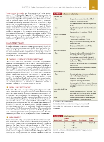
Supraventricular Tachycardia: The therapeutic approach to the manage- TABLE 36-9 Indications for Cardiac Pacing
ment of SVT is illustrated in Figure 36-7. 16,26 Vagal maneuvers (carotid
sinus massage or Valsalva maneuver) may terminate AV node reentry or Acquired AV Block
reciprocating tachycardia involving a bypass tract. Adenosine is the initial Class I Symptomatic permanent or intermittent AV block
drug of choice for regular narrow complex SVT although β-blockers, Symptomatic second degree AV block
verapamil, and diltiazem are also effective. Oral antiarrhythmic drug
therapy may be required to prevent recurrent SVT (Table 36-4). Infre- Atrial flutter or fibrillation with advanced symptomatic
quently, synchronized electrical cardioversion may be required. Catheter AV block
ablation is an effective cure for AV node reentrant tachycardias, acces- Class II Asymptomatic complete AV block with ventricular rate
sory pathways or atrial tachycardias. Multifocal atrial tachycardias may <40 bpm
58
be difficult to suppress or to achieve rate control pharmacologically and Asymptomatic type II second degree AV block
then main goal is treatment of the underlying condition (such as COPD).
Implantation of a ventricular pacemaker followed by a total AV junction Post Myocardial Infarction
ablation is may be an effective treatment option, for atrial tachycardias Class I Persistent complete heart block
unresponsive to pharmacologic treatment. 54 Persistent type II second-degree AV block
Class II Newly acquired BBB with transient high grade AV or
BRADYARRHYTHMIAS complete heart block
Disorders of impulse formation or conduction may cause bradyarrhyth- Newly acquired BBB with first degree AV block
mias. Sinus node dysfunction characterized by sinus bradycardia, sino- Newly acquired bifascicular BBB
atrial exit block and/or sinus arrest causing symptomatic bradycardia is Chronic Bifascicular Block
the most common indication for permanent cardiac pacing. AV block,
59
either permanent or intermittent is the second most common cause for Class I Symptomatic patients with fascicular block and inter-
permanent cardiac pacing. mittent high grade AV or complete heart block
■ EVALUATION OF THE PATIENT WITH BRADYARRHYTHMIAS Symptomatic patients with bifascicular block and HV
interval prolongation (>100 ms) or block distal to the His
The initial evaluation of the patient with a documented bradyarrhythmia bundle at rates <100 bpm
should include a thorough history (if the patient is able to communicate) and Class II Symptomatic with bifascicular block and no identifiable
physical examination with a focus on detecting structural heart disease. A cause of syncope
12-lead ECG and rhythm strips documenting the bradyarrhythmia should Asymptomatic with bifascicular block and intermittent
be reviewed. If clinically appropriate, such as an elderly patient with syn- type II second degree AV block
cope, carotid sinus massage should be performed to look for carotid sinus
hypersensitivity unless contraindicated, that is, carotid bruits or prior stroke. Sinus Node Dysfunction
Cardiac hemodynamic data during the arrhythmia, if available, should Class I Sinus node dysfunction with symptoms of bradycardia
be reviewed. Any drugs likely contributing to the bradyarrhythmia with or without required drug therapy
should be identified and drug levels determined if appropriate (eg, digi- Symptomatic sinus bradycardia
talis). Laboratory tests should include cardiac enzymes (CK or troponin).
An echocardiogram should be performed to determine the presence of Symptomatic chronotropic incompetence
structural heart disease and to assess ventricular function. Class II Sinus bradycardia <40-50 bpm or asystole >3 seconds
■ GENERAL PRINCIPLES OF TREATMENT and suggested symptoms not documented to be due to
bradycardia
In the ICU patient with hemodynamically significant persistent brady- Hypersensitive Carotid Sinus
cardia, transcutaneous pacing should be commenced until a temporary Class I Recurrent syncope and asystole or heart block >3 sec-
pacemaker can be inserted. Placement of a transvenous electrode cathe- onds during carotid sinus massage or clear-cut clinical
ter can usually be accomplished at the bedside via the internal jugular or situation suggestive of a vasoinhibitory response
subclavian route using a flotation pacing catheter. Fluoroscopy may be Class II Recurrent syncope without clear clinical setting but
required for positioning the pacing electrode if adequate pacing thresh- abnormal response to carotid sinus massage
olds cannot be achieved. If the bradyarrhythmia is transient, temporary
pacing may not be required and the risk/benefit of this intervention AV, atrioventricular; BBB, bundle branch block.
needs to be considered. Any reversible causes should be identified and Class I: condition where there is general consensus that a pacing system is indicated.
corrected. Drugs contributing to bradycardia should be discontinued. Class II: condition where there is consensus that a pacing system might be beneficial.
Second-degree AV block or complete heart block following an inferior See reference 59.
myocardial infarction may not be persistent. If the bradyarrhythmia
does not resolve, permanent cardiac pacing may be required. The indi-
cations for permanent cardiac pacing are summarized in Table 36-9. 59
■ PACING MODALITIES TABLE 36-10 Pacing System Code
Pacemakers started being developed in the 1960s to treat bradyarrhyth- Chamber Paced Chamber Response Rate Adaptive Multisite
Sensed
Pacing
mias. As pacemakers became more sophisticated in pacing, sensing, and
other features, codes started being developed to describe pacemaker A A O / I / T O / R O / A / V / D
function. The Three-Position code was developed in 1974. The first V V O / I / T
60
letter describes the chamber paced, the second the chamber sensed and D D O / I / T / D
the third describes how the chamber responds to a paced or intrinsic
event. Various changes have been made to the coding since then. The A, atrium; D, dual chamber; I, inhibited; O, off; R, rate modulation; T, triggered; V, ventricle.
currently used coding system still uses the same definitions for the first See references 60 and 61.
section03.indd 289 1/23/2015 2:07:14 PM