
Page 421 - Hall et al (2015) Principles of Critical Care-McGraw-Hill
P. 421
CHAPTER 36: Cardiac Arrhythmias, Pacing, Cardioversion, and Defibrillation in the Critical Care Setting 291
A
I aVR v1 v4
II aVL v2 v5
III aVF v3 v6
II
B
I aVR V1 V4
II aVL V2 V5
III aVF V3 V6
II
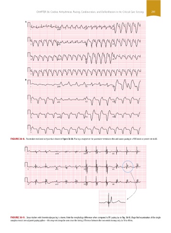
FIGURE 36-8. Pacemaker-mediated tachycardia is shown in Figure 36-8A. Placing a magnet on the pacemaker terminates this and causes pacing in a VOO mode at preset rate in 8B.
I aVR V1 V4
II aVL V2 V5
III aVF V3 V6
FIGURE 36-9. Sinus rhythm with biventricular pacing is shown. Note the morphology difference when compared to RV pacing (as in Fig. 36-8). Magnified examination of the single
complex reveals two adjacent pacing spikes—this may not always be seen since the timing difference between the two ventricles may only be 10 to 40 ms.
section03.indd 291 1/23/2015 2:07:15 PM