
Page 483 - Hall et al (2015) Principles of Critical Care-McGraw-Hill
P. 483
CHAPTER 41: Valvular Heart Disease 353
rotating inside of the valvular housing. Mechanical prostheses have a sudden onset of pulmonary edema for left-sided valves or acute decom-
clear durability advantage, but require life-long anticoagulation. pensated right heart failure and acute congestive hepatopathy (“shock
Biological valves are composed at least in part of biologic tissue, have liver”) for right-sided valves. Clinical examination is challenging, and
a lower thrombogenic potential, but uniformly deteriorate due to wear the classical description of “muffled” mechanical prosthetic sounds can
and tear and immunologic foreign body reactions. Degeneration is be subtle or even absent (in the case of multiple mechanical prostheses,
accelerated in younger patients and in patients with disordered calcium of which only one is dysfunctional; obviously, bioprosthesis do not have
metabolism. Evidence of degeneration can usually be detected by 5 years sharp sounds regardless of their functional status). A systolic ejection
after replacement. By 15 years, over 50% of tissue valves will have murmur (aortic prosthesis) or diastolic rumble (mitral and tricuspid
failed. 37,38 Fortunately, valve failure is rarely sudden, and a second opera- prosthesis) can be heard. Tachycardia, third and fourth heart sounds, as
tion can frequently be done on an elective basis. Sudden cuspal tears can well as signs of cardiogenic shock may be present.
present as an acute regurgitant lesion. Cues for diagnosis are provided by history and clinical presentation.
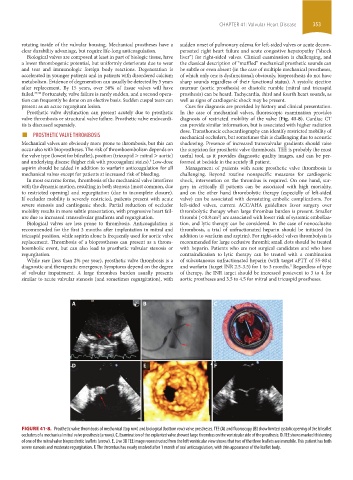
Prosthetic valve dysfunction can present acutely due to prosthetic In the case of mechanical valves, fluoroscopic examination provides
valve thrombosis or structural valve failure. Prosthetic valve endocardi- diagnosis of restricted mobility of the valve (Fig. 41-8). Cardiac CT
tis is discussed separately. can provide similar information, but is associated with higher radiation
■ PROSTHETIC VALVE THROMBOSIS dose. Transthoracic echocardiography can identify restricted mobility of
mechanical occluders, but sometimes this is challenging due to acoustic
Mechanical valves are obviously more prone to thrombosis, but this can shadowing. Presence of increased transvalvular gradients should raise
occur also with bioprostheses. The risk of thromboembolism depends on the suspicion for prosthetic valve thrombosis. TEE is probably the most
the valve type (lowest for bileaflet), position (tricuspid > mitral > aortic) useful tool, as it provides diagnostic quality images, and can be per-
and underlying disease (higher risk with procoagulant states). Low-dose formed at bedside in the acutely ill patient.
5
aspirin should be added in addition to warfarin anticoagulation for all Management of patients with acute prosthetic valve thrombosis is
mechanical valves except for patients at increased risk of bleeding. challenging. Beyond routine nonspecific measures for cardiogenic
In most extreme forms, thrombosis of the mechanical valve interferes shock, intervention on the thrombus is required. On one hand, sur-
with the dynamic motion, resulting in both stenosis (most common, due gery in critically ill patients can be associated with high mortality,
to restricted opening) and regurgitation (due to incomplete closure). and on the other hand thrombolytic therapy (especially of left-sided
If occluder mobility is severely restricted, patients present with acute valve) can be associated with devastating embolic complications. For
severe stenosis and cardiogenic shock. Partial reduction of occluder left-sided valves, current ACC/AHA guidelines favor surgery over
mobility results in more subtle presentation, with progressive heart fail- thrombolytic therapy when large thrombus burden is present. Smaller
ure due to increased transvalvular gradients and regurgitation. thrombi (<0.8 cm ) are associated with lower risk of systemic emboliza-
2
Biological valves are less prone to thrombosis. Anticoagulation is tion, and lytic therapy can be considered. In the case of nonocclusive
recommended for the first 3 months after implantation in mitral and thrombosis, a trial of unfractionated heparin should be initiated (in
tricuspid position, while aspirin alone is frequently used for aortic valve addition to warfarin and aspirin). For right-sided valves thrombolysis is
replacement. Thrombosis of a bioprostheses can present as a throm- recommended for large occlusive thrombi; small clots should be treated
boembolic event, but can also lead to prosthetic valvular stenosis or with heparin. Patients who are not surgical candidates and who have
regurgitation. contraindication to lytic therapy can be treated with a combination
While rare (less than 2% per year), prosthetic valve thrombosis is a of subcutaneous unfractionated heparin (with target aPTT of 55-80 s)
diagnostic and therapeutic emergency. Symptoms depend on the degree and warfarin (target INR 2.5-3.5) for 1 to 3 months. Regardless of type
5
of valvular impairment. A large thrombus burden usually presents of therapy, the INR target should be increased postevent to 3 to 4 for
similar to acute valvular stenosis (and sometimes regurgitation), with aortic prostheses and 3.5 to 4.5 for mitral and tricuspid prostheses.
A B C
D E F
FIGURE 41-8. Prosthetic valve thrombosis of mechanical (top row) and biological (bottom row) valve prostheses. TEE (A) and fluoroscopy (B) show limited systolic opening of the bileaflet
occluders of a mechanical mitral valve prosthesis (arrows). C. Examination of the explanted valve showed large thrombus on the ventricular side of the prosthesis. D. TEE shows marked thickening
of one of the mitral valve bioprosthetic leaflets (arrow). E. Live 3D TEE image reconstructed from the left ventricular view shows that two of the three leaflets are immobile. This patient has both
severe stenosis and moderate regurgitation. F. The thrombus has nearly resolved after 1 month of oral anticoagulation, with thin appearance of the leaflet body.
section03.indd 353 1/23/2015 2:08:03 PM