
Page 479 - Hall et al (2015) Principles of Critical Care-McGraw-Hill
P. 479
CHAPTER 41: Valvular Heart Disease 349
right ventricular function. Heavily calcified valves may obscure pres-
ence of mitral regurgitation due to acoustic shadowing of the left atrium.
Whenever in doubt, presence of concomitant mitral regurgitation
should be assessed with TEE; this will also allow assessment of the left
atrial appendage for the presence of left atrial thrombus.
Given the reliability of echocardiographic techniques, cardiac cath-
eterization is seldom used as a diagnostic tool. Mitral valve area can be
calculated by the Gorlin formula, and right heart pressures are directly
measured. Left atrial pressure can be directly measured by transseptal
puncture, and is preferred to pulmonary capillary wedge pressure (the
latter can overestimate left atrial pressure, and hence the severity of MS).
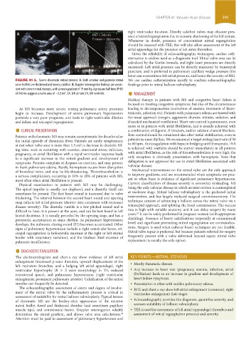
FIGURE 41-5. Severe rheumatic mitral stenosis. A. Both anterior and posterior mitral We use cardiac catheterization mostly to confirm echocardiographic
valve leaflets are thickened and heavily calcified. B. Doppler interrogation findings are consis- findings prior to mitral balloon valvuloplasty.
tent with severe mitral stenosis, with a mean gradient of 19 mm Hg. A pressure half time (PHT)
of 240 ms suggests a valve area of ~0.9 cm . LA, left atrium; LV, left ventricle. ■ MANAGEMENT
2
Medical therapy in patients with MS and congestive heart failure is
focused on treating congestive symptoms, but also of the circumstances
As MS becomes more severe, resting pulmonary artery pressures leading to decompensation (correction of anemia, treatment of thyro-
begin to increase. Development of severe pulmonary hypertension toxicosis, infection etc). Patients with pulmonary edema are treated with
portends a very poor prognosis, and leads to right ventricular dilation the usual approach (oxygen, aggressive diuresis, nitrates, sedation, and
and failure and tricuspid regurgitation. if needed mechanical ventilation). Heart rate control is paramount, even
■ CLINICAL PRESENTATION more so in patients with atrial fibrillation, and is usually achieved with
a combination of digoxin, β-blockers, and/or calcium channel blockers.
Patients with rheumatic MS may remain asymptomatic for decades after Rate control should be considered also after initial stabilization, even in
the initial episode of rheumatic fever. Patients are rarely symptomatic patients in sinus rhythm. We recommend a target resting heart rate of 50
2
at rest when valve area is more than 1.5 cm ; a decrease in diastolic fill- to 60 bpm. Anticoagulation with heparin bridging until therapeutic INR
ing time, such as occurring with exercise, emotional stress, infection, is achieved with warfarin should be started immediately in all patients
pregnancy, or atrial fibrillation with a rapid ventricular response leads with atrial fibrillation, as the risk of thromboembolism is very high; the
to a significant increase in the mitral gradient and development of only exception is obviously presentation with hemoptysis. Note that
symptoms. Patients complain of dyspnea on exertion, and may present dabigatran is not approved for use in atrial fibrillation associated with
in frank pulmonary edema. Rarely, hemoptysis occurs from disruption valvular disease.
of bronchial veins, and may be life-threatening. Thromboembolism is Mechanical interventions on the mitral valve are the only approach
a serious complication, occurring in 10% to 20% of patients with MS, to improve gradients, and are recommended when symptoms are pres-
most often when atrial fibrillation is present. ent or when there is evidence of significant pulmonary hypertension.
Physical examination in patients with MS may be challenging. Current classification of disease severity is somewhat misleading, MS
The apical impulse is usually not displaced, and a diastolic thrill can being the only valvular disease in which an intervention is contemplated
sometimes be present. The first heart sound is increased due to valve at moderate stage. Mitral balloon valvuloplasty is the preferred initial
thickening. The interval between the second heart sound and opening intervention, and has largely replaced surgical commissurotomy. The
snap reflects left atrial pressure (shorter time consistent with increased technique consists of advancing a balloon across the mitral valve via a
disease severity). The diastolic rumble is a low-pitched sound and is transseptal approach, and splitting the fused commissures. The success
difficult to hear; it is present at the apex, and can be best heard in left rate is high with suitable anatomy, and it can delay surgery by many
29
lateral decubitus. It is usually preceded by the opening snap, and has a years. It can be safely performed in pregnant women (with appropriate
presystolic accentuation in sinus rhythm. As pulmonary hypertension shielding). Presence of heavy calcifications (especially at commissural
develops, the pulmonic closing sound (P2) becomes accentuated. Other level) and significant preexisting mitral regurgitation are contraindica-
signs of pulmonary hypertension include a right ventricular heave, tri- tions. Surgery is used when catheter-based techniques are not feasible.
cuspid regurgitation (a holosystolic murmur at the right or left sternal Mitral valve repair is preferred, but because patients referred for surgery
border with respiratory variation), and the Graham Steel murmur of frequently present with a valve deformed beyond repair, mitral valve
pulmonic insufficiency. replacement is usually the only option.
■ DIAGNOSTIC EVALUATION
The electrocardiogram and chest-x ray show evidence of left atrial KEY POINTS—MITRAL STENOSIS
enlargement (increased p-wave duration, upward displacement of the
left mainstem bronchus, and a bulging left atrial appendage), right • Mostly rheumatic disease.
ventricular hypertrophy (R > S wave morphology in V1; reduced • Any increase in heart rate (pregnancy, anemia, infection, atrial
retrosternal space), and pulmonary hypertension (right ventricular fibrillation) leads to an increase in gradient and development of
enlargement, prominent pulmonary arteries). Calcification of the mitral heart failure symptoms.
annulus can frequently be detected. • Presentation is often with sudden pulmonary edema.
The echocardiographic assessment of extent and degree of involve- • ECG and chest x-ray show left atrial enlargement (common), right
ment of the mitral valve by the inflammatory process is critical in ventricular enlargement (late stage).
assessment of suitability for mitral balloon valvuloplasty. Typical lesions
of rheumatic MS are the hockey-stick appearance of the anterior • Echocardiography provides the diagnosis, quantifies severity, and
mitral leaflet, fused and thickened chordae (and sometimes papillary assesses suitability of balloon valvuloplasty.
muscle tips), and commissural fusion. Doppler interrogation reliably • TEE is used for assessment of left atrial (appendage) thrombus and
determines the mitral gradient, and allows valve area calculations. assessment of mitral regurgitation presence and severity.
28
Attention must be paid to assessment of pulmonary hypertension and
section03.indd 349 1/23/2015 2:07:51 PM