
Page 482 - Hall et al (2015) Principles of Critical Care-McGraw-Hill
P. 482
352 PART 3: Cardiovascular Disorders
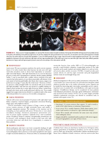
A B C
RV
RV LV
LV
RA LA
RA
D E F
FIGURE 41-7. Various causes of tricuspid regurgitation. A. Functional TR is the most common tricuspid valve disease. Note traction of the leaflets with large area of malcoaptation (arrows)
in this patient with underlying severe pulmonary hypertension. B. Color flow imaging in the same patient demonstrates massive TR. C. An uncommon cause of tricuspid regurgitation is tricuspid
valvectomy. This patient underwent tricuspid valve implantation 17 years after initial operation. D. Typical dagger shape of torrential tricuspid regurgitant signal (arrows). This is due to rapid
equalization of pressures in the right ventricle and right atrium with very large regurgitant orifice. E. This mobile mass (arrow) was seen in the right atrium shortly after a difficult pacemaker
lead extraction. Surgery confirmed ruptured papillary muscle (arrow) with severe prolapse of the entire anterior leaflet (F).
■ PATHOPHYSIOLOGY ventricular function than cardiac MRI or CT, echocardiography can
Acute severe TR is an uncommon condition, but can be seen in traumatic provide a rapid bedside evaluation. Longitudinal motion of the right
injury of the valve (mostly blunt chest trauma), but also seen with right ventricle can be easily tracked, and indexes related to it, such as tricus-
ventricular procedures (such as device lead implantation or extraction, pid annulus plane systolic excursion (TAPSE), tricuspid annulus peak
right ventricular biopsy), with rapid destruction of the valve by infectious systolic velocity (s’), and free right ventricular wall peak longitudinal
process, or rarely due to spontaneous or traumatic chordal rupture with flail systolic strain are increasingly being used.
ventricle, but in the majority of cases this is remarkably well tolerated. In an ■ MANAGEMENT
leaflets. It may be associated with a sudden volume overload of the right
early report of flail tricuspid leaflets after blunt chest trauma, surgery was Medical management is the norm in initial presentation with severe TR.
performed on average 17 years after the initial event. However, at follow- Loop diuretics are commonly used, but spironolactone should be added
35
up, presence of flail tricuspid leaflet was associated with excess mortality whenever possible due to the relative aldosterone excess. Digoxin can
and high morbidity, and there are case reports of a rapidly evolving car- be helpful for improving right ventricular contractility and for control-
36
diogenic shock picture due to acute right ventricular failure. Furthermore, ling heart rates in patients with atrial fibrillation with rapid ventricular
tricuspid valve repair can be usually performed with low risk. These results response. Ultrafiltration therapy can be considered. In those patients
suggest that surgical intervention should be considered early in the course with fulminant presentation, inotropic agents (dobutamine, milrinone)
of the disease before the occurrence of irreversible consequences. and mechanical assist devices (RVAD) can be used. Surgery provides
■ CLINICAL PRESENTATION definitive anatomical correction, and should be considered early in
patients with organic severe TR.
Patients with severe decompensated TR present with right-sided heart
failure symptoms (marked fatigue, postprandial abdominal bloating, KEY POINTS—TRICUSPID REGURGITATION
weight gain, and peripheral edema). • Functional TR is more common than organic TR, and is mostly a
Clinical examination reveals presence of large v waves on jugular result of left-sided disease or pulmonary hypertension.
venous contour, presence of right ventricular heave, and usually a
tricuspid regurgitant murmur. The intensity of the murmur can be • Acute severe TR is usually traumatic (blunt chest trauma, pace-
misleading, as many patients with severe TR have low or even absent maker lead insertion or extraction, right ventricular biopsy).
auscultatory findings. Abdominal examination demonstrates pres- • Severe TR is usually well tolerated, but some cases may present
ence of systolic hepatic expansion (“pulsatile liver”) and occasionally with cardiogenic shock.
ascites. Peripheral edema is common. Exceptionally, presentation can • Surgery should be considered early in patients with severe organic
be dramatic, with cardiogenic shock (similar to that seen with right tricuspid regurgitation and signs of heart failure.
ventricular infarction) and shock liver. Coagulopathy is common in
these patients.
■ DIAGNOSTIC EVALUATION PROSTHETIC VALVE DYSFUNCTION
Electrocardiogram and chest x-ray show nonspecific findings of right Prosthetic heart valves are classified as mechanical or biological valves.
ventricular hypertrophy, right ventricular strain, and pleural effusions. Various generations of mechanical prostheses can be seen in clinical
Echocardiography shows presence of tricuspid regurgitation and degree practice (ball-cage, tilting disk, and bileaflet). Modern mechanical pros-
of right ventricular dysfunction. While less robust in assessment of right theses have a bileaflet occluder structure, with two semicircular discs
section03.indd 352 1/23/2015 2:08:00 PM