
Page 486 - Hall et al (2015) Principles of Critical Care-McGraw-Hill
P. 486
356 PART 3: Cardiovascular Disorders
• Malouf J, Le Tourneau T, Pellikka P, et al. Aortic valve stenosis in INTRODUCTION
community medical practice: determinants of outcome and impli- Aortic dissection occurs much more frequently than previously appreci-
cations for aortic valve replacement. J Thorac Cardiovasc Surg. ated and is actually the most common catastrophe affecting the aorta,
2012;144(6):1421-1427. occurring 2 to 3 times more commonly than acute abdominal aortic
• Nishimura RA, Grantham JA, Connolly HM, Schaff HV, Higano aneurysm rupture. Although the diagnosis is sometimes obvious, the
1-3
ST, Holmes DR Jr. Low-output, low-gradient aortic stenosis in majority of cases are not clear-cut and the patient’s survival will depend
patients with depressed left ventricular systolic function: the on a high index of suspicion by the physician despite a myriad of differ-
clinical utility of the dobutamine challenge in the catheterization ent clinical presentations. Time is of the essence as the mortality is 50%
laboratory. Circulation. 2002;106(7):809-813. for the first 48 hours without treatment and 85% to 90% over 3 months.
• Nishimura RA, Rihal CS, Tajik AJ, Holmes DR Jr. Accurate mea- The typically hypertensive patient must have their blood pressure and
surement of the transmitral gradient in patients with mitral steno- pain controlled quickly followed by rapid diagnosis with definitive
sis: a simultaneous catheterization and Doppler echocardiographic imaging and immediate relegation to the appropriate therapy of either
study. J Am Coll Cardiol. July 1994;24(1):152-158. emergency surgery or medical management/endostenting.
• Nkomo VT, Gardin JM, Skelton TN, Gottdiener JS, Scott CG,
Enriquez-Sarano M. Burden of valvular heart diseases: a popula- PATHOGENESIS
tion-based study. Lancet. 2006;16;368(9540):1005-1011. Previously, aortic dissections were referred to as dissecting aneurysms, as
• Wilson W, Taubert KA, Gewitz M, et al. Prevention of infective originally coined by Laënnec. This is a misnomer in that the pathology is a
endocarditis: guidelines from the American Heart Association: a dissecting hematoma that separates the intima and inner layers of the media
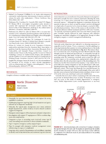
guideline from the American Heart Association Rheumatic Fever, from the outer medial and adventitial layers (Fig. 42-1). The intima is there-
Endocarditis, and Kawasaki Disease Committee, Council on fore not aneurysmal, and is, if anything, narrowed. Blood invades the media
Cardiovascular Disease in the Young, and the Council on Clinical through a tear in the intima and proceeds ante- or retrogradely through the
Cardiology, Council on Cardiovascular Surgery and Anesthesia, aortic wall, forming a false lumen. In type A dissections (originating in the
4
and the Quality of Care and Outcomes Research Interdisciplinary ascending aorta) the hematoma commonly spirals around the right and
Working Group. Circulation. October 9, 2007;116(15):1736-1754. posterior aspects of the ascending aorta, supraposteriorly along the arch,
• Zoghbi WA, Enriquez-Sarano M, Foster E, et al. Recommendations and then down the left and posterior aspects of the descending aorta. The
for evaluation of the severity of native valvular regurgitation hematoma may then have several serious sequelae. It may rupture into the
with two-dimensional and Doppler echocardiography. J Am Soc pericardial space causing tamponade or into the pleural space with exsan-
Echocardiography. 2003;16(7):777-802. guinating hemorrhage, especially in type B dissections (begin after the left
subclavian artery). This occurs less frequently than expected because the
adventitial layer represents 66% of the overall strength of the aortic wall. It
REFERENCES may also cause occlusion of aortic branch arteries or prolapse of one or more
of the aortic valve cusps, resulting in acute aortic insufficiency.
Complete references available online at www.mhprofessional.com/hall Generally the tear is due to either a weakening of the wall of the aorta,
an increase in luminal shear stress, or both. Weakening of the aortic
wall can occur as the result of medial degeneration or iatrogenic
Aortic Dissection injury. Medial degeneration (cystic medial degeneration or necrosis)
CHAPTER
42 Vlad Cotarlan Left common
Joseph J. Austin
Innominate carotid artery
artery
Left subclavian
artery
KEY POINTS
• Potentially the most important diagnosis with highest life-saving 4
capability in medicine.
• Challenging diagnosis requiring high clinical suspicion and quick,
efficient use of diagnostic modalities.
• Clinically, the typical pain, incongruous poor tissue perfusion
despite hypertension, and/or evidence of aortic branch occlusion 2
suggest the diagnosis. 1
• Emergent control/support of blood pressure and pain is imperative.
• Investigation with urgent CT angiogram or TEE to confirm diag-
nosis and complications.
• Categorize as type A (ascending aorta involved) versus type B Aortic
(only descending aorta involved) to direct definitive treatment. valve
• Type A requires emergency cardiac surgical repair. 3
• Type B managed with emergency medical management versus
endo stenting or surgery if complicated. FIGURE 42-1. Aortic dissection begins with an intimal tear (1) leading to a hematoma
that separates the layers of the aortic wall. The sequelae are rupture through the adventitia
• Long-term strict control of hypertension and surveillance important into the pericardium (2), prolapse of the aortic valve cusps leading to aortic insufficiency
to identify need for late intervention and maximize long-term survival. (3), compression of the aortic branch vessels (4), and aneurysmal dilation of the ascending
arch and descending aorta.
section03.indd 356 1/23/2015 2:08:10 PM