
Page 485 - Hall et al (2015) Principles of Critical Care-McGraw-Hill
P. 485
CHAPTER 41: Valvular Heart Disease 355
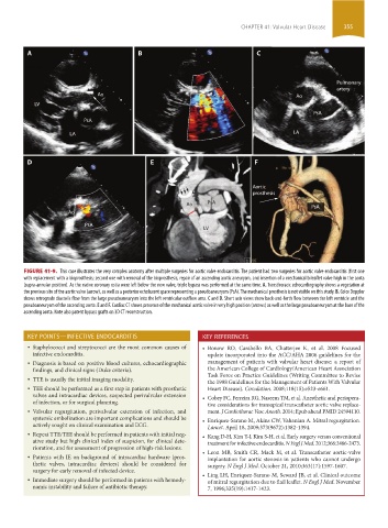
A B C
Pulmonary
artery
Ao Ao
LV
PsA
PsA
LA LA
D E F
Aortic
prosthesis
Ao PsA PsA
Ao
PsA
LV
FIGURE 41-9. This case illustrates the very complex anatomy after multiple surgeries for aortic valve endocarditis. The patient had two surgeries for aortic valve endocarditis (first one
with replacement with a bioprosthesis; second one with removal of the bioprosthesis, repair of an ascending aortic aneurysm, and insertion of a mechanical bileaflet valve high in the aorta
(supra-annular position). As the native coronary ostia were left below the new valve, triple bypass was performed at the same time. A. Transthoracic echocardiography shows a vegetation at
the previous site of the aortic valve (arrow), as well as a posterior echolucent space representing a pseudoaneurysm (PsA). The mechanical prosthesis is not visible on this study. B. Color Doppler
shows retrograde diastolic flow from the large pseudoaneurysm into the left ventricular outflow area. C and D. Short axis views show back-and-forth flow between the left ventricle and the
pseudoaneurysm of the ascending aorta. E and F. Cardiac CT shows presence of the mechanical aortic valve in very high position (arrows) as well as the large pseudoaneurysm at the base of the
ascending aorta. Note also patent bypass grafts on 3D CT reconstruction.
KEY POINTS—INFECTIVE ENDOCARDITIS KEY REFERENCES
• Staphylococci and streptococci are the most common causes of • Bonow RO, Carabello BA, Chatterjee K, et al. 2008 Focused
infective endocarditis. update incorporated into the ACC/AHA 2006 guidelines for the
• Diagnosis is based on positive blood cultures, echocardiographic management of patients with valvular heart disease: a report of
findings, and clinical signs (Duke criteria). the American College of Cardiology/American Heart Association
Task Force on Practice Guidelines (Writing Committee to Revise
• TTE is usually the initial imaging modality.
the 1998 Guidelines for the Management of Patients With Valvular
• TEE should be performed as a first step in patients with prosthetic Heart Disease). Circulation. 2008;118(15):e523-e661.
valves and intracardiac devices, suspected perivalvular extension • Cobey FC, Ferreira RG, Naseem TM, et al. Anesthetic and periopera-
of infection, or for surgical planning. tive considerations for transapical transcatheter aortic valve replace-
• Valvular regurgitation, perivalvular extension of infection, and ment. J Cardiothorac Vasc Anesth. 2014; Epub ahead PMID 24594110.
systemic embolization are important complications and should be • Enriquez-Sarano M, Akins CW, Vahanian A. Mitral regurgitation.
actively sought on clinical examination and ECG. Lancet. April 18, 2009;373(9672):1382-1394.
• Repeat TTE/TEE should be performed in patients with initial neg- • Kang D-H, Kim Y-J, Kim S-H, et al. Early surgery versus conventional
ative study but high clinical index of suspicion, for clinical dete- treatment for infective endocarditis. N Engl J Med. 2012;366:2466-2473.
rioration, and for assessment of progression of high-risk lesions.
• Patients with IE on background of intracardiac hardware (pros- • Leon MB, Smith CR, Mack M, et al. Transcatheter aortic-valve
implantation for aortic stenosis in patients who cannot undergo
thetic valves, intracardiac devices) should be considered for surgery. N Engl J Med. October 21, 2010;363(17):1597-1607.
surgery for early removal of infected device.
• Immediate surgery should be performed in patients with hemody- • Ling LH, Enriquez-Sarano M, Seward JB, et al. Clinical outcome
of mitral regurgitation due to flail leaflet. N Engl J Med. November
namic instability and failure of antibiotic therapy. 7, 1996;335(19):1417-1423.
section03.indd 355 1/23/2015 2:08:09 PM