
Page 480 - Hall et al (2015) Principles of Critical Care-McGraw-Hill
P. 480
350 PART 3: Cardiovascular Disorders
large v waves are present on pulmonary artery wedge pressure tracing.
• Cardiac catheterization confirms the diagnosis, but is mostly used Cardiac output can be dramatically reduced, as the left ventricle ejects
for balloon valvuloplasty. into the left atrium, leading to cardiogenic shock. Often surgery has to
• Intensive care unit management includes oxygenation, diuretics, nitrates, be performed emergently.
mechanical ventilation to manage high-pressure pulmonary edema. In chronic MR, the left ventricle progressively remodels, with char-
• Percutaneous balloon mitral valvuloplasty is the preferred initial acteristic enlargement. Since afterload is reduced (it is a combination
of aortic and left atrial pressure) ejection indexes are initially increased.
treatment with results equivalent to surgical commissurotomy. A reduction toward “normal” values heralds impairment of myocardial
Can be done in pregnant women with low risk. function. Indeed, an ejection fraction of less than 60% and left ven-
• Surgical treatment: mostly in calcified valves or with MR requir- tricular end-systolic diameter of more than 40 mm are markers of left
ing valve replacement. ventricular dysfunction in patients with severe chronic MR, and are
associated with poor long-term outcome. Ejection fraction less than
32
40% represents advanced cardiac dysfunction and indicates poor post-
operative outcome. Chronic MR leads to left atrial enlargement, which
33
can be massive. Pulmonary hypertension develops in late stages of the
MITRAL REGURGITATION disease. Atrial fibrillation is very frequent.
■ ETIOLOGY ■ CLINICAL PRESENTATION
Mitral regurgitation (MR) is caused by either a disease of the leaflets and/ The symptoms of patients with MR depend on the acuity and severity
or subvalvular apparatus (organic, nonischemic MR) or malcoaptation of the disease. In patients with chronic MR, symptoms develop in older
of an otherwise normal mitral valve due to tethering (functional and patients often with signs of diastolic left ventricular dysfunction. As with
ischemic MR). The most common cause of chronic MR in the Western all other chronic valvular diseases, a concomitant illness precipitates
30
world is myxomatous degeneration, occurring in isolation (primary) or cardiac decompensation on the background of reduced cardiac reserve.
in association with other connective tissue disease (secondary; Marfan Patients present with symptoms and signs of pulmonary congestion;
and Ehlers-Danlos syndromes, osteogenesis imperfecta, and pseudoxan- in the case of long-standing disease, signs of right ventricular failure
thoma elasticum). The leaflets and chordae are thickened and elongated, may also be present (peripheral edema, hepatic congestion). The apical
leading to mitral valve prolapse and regurgitation. At the other end of impulse is usually displaced, and a palpable early diastolic filling impulse
the spectrum, functional/ischemic MR is a disease of the ventricle rather may be present (palpable S3). The MR murmur is holosystolic, high
than of the valve. Malcoaptation is caused in this case by traction on the pitched, and loudest at the apex. Radiation is classically to the axilla, but
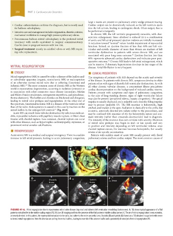
mitral leaflets by the enlarged left ventricle (Fig. 41-6). eccentric, anteriorly directed jets radiate rather to the precordium, and
31
Acute MR can result from valvular destruction by infectious endocar- may be confused with murmur of AS; the holosystolic nature and con-
ditis, myocardial ischemia with papillary muscle rupture, or blunt chest stant intensity (rather than crescendo-decrescendo) lead to diagnosis.
trauma with chordal rupture. Less common, chordal rupture can occur The intensity of the murmur does not correlate with severity. Murmurs
with other diseases, such as hypertrophic cardiomyopathy, myxomas, or of mitral valve prolapse may begin in mid- or late systole and vary
prominent mitral annular calcifications. in position and intensity depending on left ventricular volume; once
■ PATHOPHYSIOLOGY chordal rupture ensues, the murmur becomes holosystolic, but usually
retains a late systolic accentuation.
Acute severe MR is a medical and surgical emergency. There is a sudden Patients with sudden onset of acute MR usually present with florid
increase in left atrial pressure, leading to acute pulmonary congestion; pulmonary edema and low cardiac output. The most common causes of
A LA B C
Flail Flail
LV
D E F
Diastolic Systolic
frame frame
LA
LA
Tethering
LV LV
FIGURE 41-6. Mitral regurgitation due to myxomatous mitral valve disease (top row) and ischemic left ventricular remodeling (bottom row). A. TEE shows typical appearance of a flail
posterior mitral leaflet in the middle scallop region (P2). B. Live 3D imaging confirms the presence of the flail posterior middle scallop (arrow). C. The jet of mitral regurgitation is very eccentric,
anteriorly directed. In this patient, the murmur did not radiate to the axilla, but rather to the entire precordial area. Diastolic (D) and systolic (E) frames on a TEE obtained in a patient with severe
ischemic mitral regurgitation. Note the chordae are pulling the mitral leaflets, leading to override of the anterior leaflet and a posteriorly directed jet of mitral regurgitation (F).
section03.indd 350 1/23/2015 2:07:56 PM