
Page 552 - Hall et al (2015) Principles of Critical Care-McGraw-Hill
P. 552
372 PART 4: Pulmonary Disorders
for generation of high-energy adenosine triphosphate (ATP) bonds
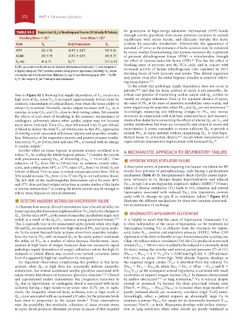
TABLE 43-2 Oxygen Cost (V ˙ ) of Breathing and Fever in 20 Critically III Patients through aerobic glycolysis; then excess pyruvate circulates in normal
O 2
Breathing (Mean ± SD) 15 Fever (Mean ± SD) 17 equilibrium with excess lactate, and clinicians mistake this lactic
Mode V ˙ Body Temperature V ˙ acidosis for anaerobic metabolism. Alternatively, the appearance of
O 2
O 2
elevated L/P ratios in the presence of lactic acidosis may be explained
CPAP 255 ± 92 39.4°C ± 0.8°C 359 ± 65 by recent studies demonstrating that hypoxia increases the expression
AC/MR 209 ± 79 37.0°C ± 0.5°C 295 ± 57 of pyruvate dehydrogenase kinase (PDK) in mitochondria through
24
V ˙ resp 46 ± 21 V ˙ /°C 27.5 ± 8.1 the effect of hypoxia-inducible factor (HIF). This has the effect of
O 2
O 2
AC/MR, assist-control mode with muscle relaxation (full mechanical ventilation); °C, body temperature blocking entry of pyruvate into the TCA cycle, and in concert with
increased activity of lactate dehydrogenase (also regulated by HIF)
in degrees centigrade; CPAP, continuous positive airway pressure (spontaneous breathing); V ˙ , oxygen elevating levels of both pyruvate and lactate. This altered regulation
O 2
consumption (mL/min) by spirometer (Deltatrac); V ˙ resp, O2 cost of breathing equals CPAP − AC/MR; may persist even after the initial hypoxic stimulus is removed follow-
O 2
V ˙ /°C, the change in V ˙ per °C change in body temperature.
ing resuscitation. 24,25
O 2
O 2
To the extent that pathologic supply dependence does not occur in
patients, 26,27 and that the lactic acidosis of sepsis is not anaerobic, the
lines in Figure 43-1 showing that supply dependence of V ˙ occurs at a critical care practice of maximizing cardiac output and Q ˙ confers no
O 2
O 2
high level of Q ˙ when V ˙ is increased (approximately 450 mL/min) by benefit on oxygen utilization. Even in the apparent absence of sepsis,
O 2
–
O 2
common concomitants of critical illness, even when the tissue ability to the value of Pv O at the onset of anaerobic metabolism varies widely, and
–
2
28
extract O 2 is normal. Normally, cardiac output increases with V ˙ , as in some organs may be anaerobic when Pv O 2 and Q ˙ are not worrisome.
O 2
O 2
– – close to their resting values. Yet consider – and venous
exercise, to keep Cv O 2 and Cv O 2 Accordingly, measuring and following changes in Pv O 2
the effects of such work of breathing in the common circumstance of saturation in conjunction with acid-base status and lactic acid measure-
cardiogenic pulmonary edema when cardiac output may not increase ments allow deductions concerning the effects of altered Q ˙ on V ˙ and
O 2
O 2
– aerobic metabolism, but these are nonspecific and subject to errors and
much above 5.0 L/min. Then Cv O 2 must fall toward 5 mL O 2 per 100 mL
– approaches uncertainties. It seems reasonable to ensure sufficient Q ˙ to provide a
O 2
of blood to deliver the total V ˙ of 450 mL/min so that Pv O 2 – O 2
22 mm Hg, a level associated with tissue hypoxia and anaerobic metabo- normal Pv O 2 in septic patients without maximizing Q ˙ to treat hypo-
O 2
lism. Relaxation of the respiratory muscles and positive pressure ventila- thetical tissue O 2 extraction defects, 26,27 except where measurement of
– organ acidosis demonstrates improvement with increased Q ˙ . 28
O 2
tion reduce V ˙ to 250 mL/min and raise Pv O 2 to normal with no change O 2
in cardiac output. 15
Another effect on tissue hypoxia in patients already ventilated is to
reduce V ˙ by cooling the febrile hypoxic patient. Consider the patient A MECHANISTIC APPROACH TO RESPIRATORY FAILURE
17
reduction of V ˙ from 500 to 250 mL/min by sedation, muscle relax- ■ HYPOXEMIC VERSUS VENTILATORY FAILURE
O 2
= 15 mL/dL). Then
of 40 mm Hg (Ca O 2
with pneumonia causing Pa O 2
O 2
– A descriptive survey of patients requiring mechanical ventilation for RF
ation, and cooling from 40°C to 37°C raises Cv O 2 from 5 to 10 mL O 2 per
100 mL of blood. This increase in mixed venous saturation from 25% to reveals four patterns of pathophysiology, each having a predominant
– mechanism (Table 43-3). Intrapulmonary shunt (Q ˙ s/Q ˙ t) causes hypox-
50% would increase Pv O 2 from 22 to 27 mm Hg in normothermic blood.
The left shift of the oxyhemoglobin dissociation curve between 40°C emia refractory to O 2 therapy despite hyperventilation and reduced
29
and 37°C does not limit oxygen extraction in canine studies of the limits Pa CO 2 in type I or acute hypoxemic respiratory failure (AHRF). Prim ary
of aerobic metabolism, so cooling the febrile patient may be enough to failure of alveolar ventilation (V ˙ a) leads to CO 2 retention and arterial
18
relieve tissue hypoxia in critical situations. hypercapnia associated with reduced Pa O 2 ; this hypoxemia corrects
■ DETECTING ANAEROBIC METABOLISM IN RESPIRATORY FAILURE easily with O 2 therapy in type II or ventilatory failure. Figure 43-2
30
illustrates the different mechanisms for these two common abnormali-
To illustrate how several clinical interventions have a beneficial effect on ties in pulmonary O 2 exchange.
at the onset of anaerobic metabolism might vary ■
–
tissue hypoxia, this discussion emphasized how Pv O 2 tracks the changes in
– ABNORMALITIES IN PULMONARY GAS EXCHANGE
O 2
Q ˙ . Yet the value of Pv O 2
widely as a result of the Q ˙ /V ˙ variance among peripheral tissues. 19,20 It is helpful to recall that the cause of hypercapnia (inadequate V ˙ a)
O 2
O 2
This is especially true in the resuscitated septic patient when very high is often independent of the cause of hypoxemia, so the treatment for
– and lactic acido- hypercapnia (raising V ˙ a) is different from the treatment for hypox-
Q ˙ t and Q ˙ are associated with very high values of Pv O 2
O 2
sis. To the extent that such lactic acidosis arises from anaerobic metabo- , positive end-expiratory pressure [PEEP]). When CNS
– with increased Q ˙ in the septic patient confounds emia (raise Fi O 2
depression of the drive to breathe or loss of neuromuscular coupling (see
lism, the rise in Pv O 2 O 2
– as a marker of tissue hypoxia. Furthermore, lactic Chap. 54) reduces minute ventilation (V ˙ e), the CO 2 produced at rest each
the utility of Pv O 2
acidosis at high levels of oxygen transport does not necessarily signal minute (V ˙ CO 2 = 200 mL/min) is added to the reduced V ˙ a (normally about
pathologic supply dependence of oxygen utilization; rather, the high O 2 4 L/min), raising the alveolar and arterial partial pressures (P) of CO 2
demands of critical illness may exceed even normal extraction limits (Pa CO 2 = Pa CO 2 = k × V ˙ CO 2 /V ˙ a = 0.863 mm Hg/L/mL × (200 mL/min/
from the apparently high but insufficient O 2 transport. 4.0 L/min), or about 40 mm Hg). Mild alveolar hypoxia develops as
An important observation complicating this problem is that lactic the required oxygen uptake (V ˙ ) is absorbed from the reduced V ˙ a
O 2
acidosis when Q ˙ is high does not necessarily indicate anaerobic (Pa O 2 = Pi O 2 − Pa CO 2 /R, where Pa O 2 = Fi O 2 × (Pbar − P H 2 O) and R =
O 2
metabolism, but instead accelerated aerobic glycolysis associated with V ˙ c O 2 /V ˙ ), so the consequent arterial hypoxemia is corrected with small
O 2
sepsis-related disturbance of important glycolytic enzymes. 21,22 Clinical increments in inspired oxygen fraction (Fi O 2 ). In diseases characterized
and experimental studies demonstrate that progressive reduction in by airflow obstruction 30-32 or lung restriction, V ˙ a is reduced despite
33
Q ˙ due to hypovolemic or cardiogenic shock is associated with lactic normal or increased V ˙ e because the dead space:tidal volume ratio
O 2
acidemia having a high lactate-to-pyruvate ratio (L/P); yet, in septic [Vds/V t = (Pa CO 2 − Pe CO 2 )/Pa CO 2 ] is increased when large numbers of
shock, the frequently observed lactic acidemia, even at high levels of poorly perfused alveoli are excessively ventilated (high V ˙ a/Q ˙ units).
Q ˙ , is not associated with an increased L/P ratio, for the pyruvate levels Accordingly, when a patient requires an abnormally large V ˙ e to
O 2
have risen in proportion to the lactate levels. These observations maintain a normal Pa CO 2 , the causes are an abnormally increased V ˙ CO 2 ,
23
raise the possibility that metabolic utilization of tissue protein stores increased Vds/V t, or both. Hypoxemia develops with airflow obstruc-
in septic shock produces abundant pyruvate in excess of that required tion or lung restriction when other alveoli are poorly ventilated in
section04.indd 372 1/23/2015 2:18:41 PM