
Page 555 - Hall et al (2015) Principles of Critical Care-McGraw-Hill
P. 555
CHAPTER 43: The Pathophysiology and Differential Diagnosis of Acute Respiratory Failure 375
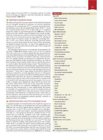
mirror image of the cause of RF (ie, a systematic approach to reverse TABLE 43-4 Liberation of the Patient From Mechanical Ventilation
factors increasing the respiratory load and decreasing the respiratory
muscle strength) (Table 43-4). Type I: AHRF
■ PERIOPERATIVE RESPIRATORY FAILURE Reduce edema production
Enhance edema clearance
The physician frequently encounters patients in the perioperative period Treat pneumonia
who are unusually susceptible to atelectasis as a primary mechanism Drain pleural effusions
causing type III or perioperative RF. 45,46 In general, abnormal abdomi-
nal mechanics reduce the end-expired lung volume (↓FRC) 47-49 below Stabilize chest wall
the increased closing volume (↑CV) in these patients, 47,50,51 leading to Minimize dead space
progressive collapse of dependent lung units (see Table 43-3). The end Type II: Airflow obstruction
result can be type I AHRF, or type II ventilatory RF, or both. Yet iden- Hypoxemia—give O
tification of atelectasis as a distinct mechanism leading to this third 2
type of RF can be harnessed to prevent lung collapse by reducing the Reverse sedation
adverse effects of common clinical circumstances promoting reduc- Bronchodilation
tion in FRC, and of those conditions promoting abnormal airways Clear bronchial secretions
closure at increased lung volume. Because many of these mechanisms
are shared by patients with type I or type II RF, implementation of Treat bronchial infection
approaches to minimize atelectasis should be a part of the management Pneumothorax—chest tube
of all patients with RF. Fractured ribs—nerve block
The principles of preventing or reversing type III perioperative RF Decrease intrinsic PEEP
are listed in Table 43-4. Bedside nurses in the ICU turn the patient Allow bicarbonate accumulation
from side to side every 1 to 2 hours; during this time, they provide vig-
orous chest physiotherapy with pummeling, chest vibration, and endo- Reduce CO production
2
tracheal suction. In patients vulnerable to atelectasis, a fourth position Correct malnutrition
30° to 45° upright is helpful by reducing the load imposed by the abdo- Type III: Perioperative respiratory failure
men; also, the addition of sighs, noninvasive ventilation (eg, CPAP or
bilevel positive airway pressure) returns the end-expired lung volume Posturize and pummel
to a position above the patient’s closing volume. Special attention to Ventilate 45° upright
50
the treatment of incisional or abdominal pain (eg, epidural anesthesia Treat incisional/abdominal pain
or transcutaneous electrical nerve stimulation) and to minimization of Drain ascites
the intra-abdominal pressure of ascites or tight bandages helps prevent Reexpand atelectasis early
atelectasis. 48,42 When lobe or lung collapse is detected by physical or
radiologic examination, an early, aggressive approach to reexpansion Stop smoking 6 weeks preoperatively
includes placing the patient in the lateral decubitus position with the Avoid overhydration
collapsed lobe uppermost for vigorous pummeling and suctioning, Type IV: Shock
and then increasing the tidal volume progressively to a pressure limit
of 40 cm H 2O with end inspiratory pauses. Reexpansion often occurs Hypoperfusion
within 10 minutes and is signaled by a fall in the Pel associated with Hypotension
the normal tidal volume at the end of the reexpansion maneuver; Anemia
if this reexpansion is not confirmed radiologically, repeating these Hypoxia
maneuvers after bronchoscopy to clear endobronchial obstructions
is reasonable. Once reexpansion has occurred, the implementation of Sepsis
increased levels of PEEP and/or sighs often prevents further episodes Fever
of atelectasis. Discontinuation of smoking at least 6 weeks prior to Acidosis
elective operations reduces bronchorrhea and atelectasis, and avoid- Electrolytes (K , Ca , Mg , PO )
53
+
2+
2 −
2+
ing overhydration in perioperative patients especially vulnerable to 4
atelectasis reduces this problem. Protein-calorie nutrition
■ HYPOPERFUSION STATES CAUSE TYPE IV RESPIRATORY FAILURE Common confounding conditions
Neuromuscular disease
A significant number of ventilated patients fall outside the categories of Muscle-relaxing drugs
type I, II, or III RF. These are the patients who have been intubated and Coma, sedation
stabilized with ventilatory support during resuscitation from a hypo-
perfusion state, so type IV RF is most commonly due to cardiogenic, Cerebrovascular accident
hypovolemic, or septic shock without associated pulmonary problems Subclinical status epilepticus
(see Chap. 31). The appropriate rationale for ventilator therapy in these Hypothyroidism
patients who are frequently tachypneic with erratic respiratory pat- Phrenic nerve paralysis
terns is to stabilize gas exchange and minimize the steal of a limited
cardiac output by the working respiratory muscles until the mechanism Respiratory muscle fatigue
for the hypoperfusion state is identified and corrected. 37,54,55 Note that Respiratory muscle exercise program
liberation from the ventilator of the patient with type IV RF is simple: Tone
When shock is corrected, the patient resumes spontaneous breathing Power
and is extubated. Note further that when patients with type I, II, or III
RF suffer a concurrent hypoperfusion state, the causes of reduced blood Coordination
flow, hypotension, anemia, acidosis, and sepsis need identification and Animation and mobilization
correction as part of the liberation process (see Table 43-4). Accordingly, AHRF, acute hypoxemic respiratory failure; PEEP, positive end-expiratory pressure.
section04.indd 375 1/23/2015 2:18:43 PM