
Page 689 - Hall et al (2015) Principles of Critical Care-McGraw-Hill
P. 689
508 PART 4: Pulmonary Disorders
is unstable or there is concern that the air leak may be pleural air leak used to create a tract that is directed diagonally from the skin over the
because of mechanical ventilation, some consensus statements recom- intercostal space, over the rib, and into the next intercostal space above
mend larger bore chest tubes (24-28 French). Malignant effusions the skin incision. Care should be taken to stay close to the top of the rib
50
should also be drained with smaller caliber chest tubes, as multiple in order to avoid injury to the neurovascular bundles that run beneath
studies have now shown they are as efficacious and more comfortable each rib. Once the parietal pleura is reached, a Kelly clamp should be
than large bore chest tubes. 24,25 Indeed, several studies have demon- used to carefully pierce this well-innervated tissue plane. Kelly clamps
strated lower pain scores, reduced analgesia requirement and increased are then used to enlarge the tract into the pleural space so that the chest
comfort when smaller caliber tubes are used regardless of indication for tube can be safely inserted into the pleural space. The operator’s index
initial placement. 24,38 Traditionally a large bore chest tube, often times finger can be used for blunt dissection to assist in accessing the parietal
≥32 French, is placed to drain hemothorax in an effort to prevent tube pleura. Once the pleural space is accessed, often there will be the release
blockage from viscous fluid and blood clot. Interestingly, a recent study of either air or pleural fluid through the tract. The index finger is placed
comparing 28 to 32 versus 36 to 40 French chest tubes in trauma patients through the tract and used to sweep fully around the insertion site in
did not identify any clinically relevant differences between the two order to ensure there are no adhesions that would prevent proper tube
groups. However, there is debate over the size of the chest tube to insert placement. Subsequently, the chest tube is inserted through the tract
34
for patients with empyema. The British guidelines support the place- into the pleural space. The proximal clamp is released once it is in the
ment of image guided small bore chest drains in patients with empyema space and removed. The chest tube is directed either to the apex to drain
even though there are variable success rates of these smaller drainage a pneumothorax or to the base of the lung to drain a pleural effusion.
systems in numerous studies. 39-45 Inadequate evacuation of empyema Once in place, the chest tube is secured to the skin with the use of a
with small bore chest tubes is the most frequent complication, but some mattress suture through the incision and around the tube. This suture
investigators argue that this can be mitigated with frequent flushing of is wrapped around the tube repeatedly and tied down multiple times to
the drainage system. While many practitioners recommend large bore ensure a secure hold. Additional interrupted sutures may be needed to fully
37
chest tubes, 28 to 36 French, as definitive treatment of empyema it is close the incision. The chest tube is connected to a pleural drainage device
likely that correct positioning of the chest tube is just as important, if not and secured. The distal clamp is released, and there should be visualization
more important, than the size of the tube. 35 of either fluid in the collecting chamber of the device or air bubbles in the
■ PLACEMENT water seal chamber. Petroleum jelly gauze is used to wrap the insertion
site of the chest tube and sterile gauze placed over this. A secure pressure
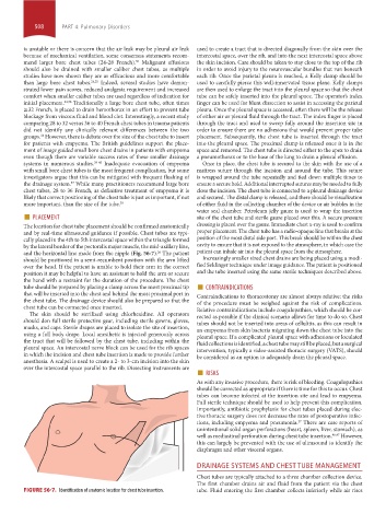
The location for chest tube placement should be confirmed anatomically dressing is placed over the gauze. Immediate chest x-ray is used to confirm
and by real-time ultrasound guidance if possible. Chest tubes are typi- proper placement. The chest tube has a radio-opaque line that breaks at the
cally placed in the 4th to 5th intercostal space within the triangle formed position of the most distal side port. This break should be within the chest
by the lateral border of the pectoralis major muscle, the mid-axillary line, cavity to ensure that it is not exposed to the atmosphere, in which case the
and the horizontal line made from the nipple (Fig. 56-7). The patient patient can inhale air into the pleural space from the atmosphere.
26
should be positioned in a semi-recumbent position with the arm lifted Increasingly smaller sized chest drains are being placed using a modi-
over the head. If the patient is unable to hold their arm in the correct fied Seldinger technique under image guidance. The patient is positioned
position it may be helpful to have an assistant to hold the arm or secure and the tube inserted using the same sterile techniques described above.
the hand with a restraint for the duration of the procedure. The chest
tube should be prepared by placing a clamp across the most proximal tip ■ CONTRAINDICATIONS
that will be inserted into the chest and behind the most proximal port in Contraindications to thoracostomy are almost always relative: the risks
the chest tube. The drainage device should also be prepared so that the of the procedure must be weighed against the risk of complications.
chest tube can be connected once inserted. Relative contraindications include coagulopathies, which should be cor-
The skin should be sterilized using chlorhexidine. All operators rected as possible if the clinical scenario allows for time to do so. Chest
should don full sterile protective gear, including sterile gowns, gloves, tubes should not be inserted into areas of cellulitis, as this can result in
masks, and caps. Sterile drapes are placed to isolate the site of insertion, an empyema from skin bacteria migrating down the chest tube into the
using a full body drape. Local anesthetic is injected generously across pleural space. If a complicated pleural space with adhesions or loculated
the tract that will be followed by the chest tube, including within the fluid collections is identified, a chest tube may still be placed, but a surgical
pleural space. An intercostal nerve block can be used for the rib spaces intervention, typically a video-assisted thoracic surgery (VATS), should
in which the incision and chest tube insertion is made to provide further be considered as an option to adequately drain the pleural space.
anesthesia. A scalpel is used to create a 2- to 3-cm incision into the skin
over the intercostal space parallel to the rib. Dissecting instruments are ■ RISKS
As with any invasive procedure, there is risk of bleeding. Coagulopathies
should be corrected as appropriate if there is time for this to occur. Chest
tubes can become infected at the insertion site and lead to empyema.
Full sterile technique should be used to help prevent this complication.
Importantly, antibiotic prophylaxis for chest tubes placed during elec-
tive thoracic surgery does not decrease the rates of postoperative infec-
tions, including empyema and pneumonia. There are case reports of
27
unintentional solid organ perforations (heart, spleen, liver, stomach), as
well as mediastinal perforation during chest tube insertion. 46,47 However,
this can largely be prevented with the use of ultrasound to identify the
diaphragm and other visceral organs.
DRAINAGE SYSTEMS AND CHEST TUBE MANAGEMENT
Chest tubes are typically attached to a three chamber collection device.
The first chamber drains air and fluid from the patient via the chest
FIGURE 56-7. Identification of anatomic location for chest tube insertion. tube. Fluid entering the first chamber collects inferiorly while air rises
section04.indd 508 1/23/2015 2:20:22 PM