
Page 688 - Hall et al (2015) Principles of Critical Care-McGraw-Hill
P. 688
CHAPTER 56: Thoracostomy 507
given their underlying lung disease. The presence of breathlessness or a
pneumothorax >2 cm in the setting of a secondary pneumothorax or an
inadequate response to air aspiration in a primary pneumothorax should
prompt the physician to insert a chest drain to fully evacuate pleural air. 2
■ PLEURAL EFFUSION
Chest imaging should be performed when a patient’s clinical presenta-
tion or exam is suggestive of pleural effusion. A posteroanterior chest
x-ray is often helpful in confirming the presence of pleural effusion and
a lateral x-ray can often have additional value. Chest computed tomog-
30
raphy can identify smaller effusions and is better able to delineate the
characteristics of a complicated effusion. Increasingly, ultrasonography
is used to localize pleural fluid, quantify its size, and guide sampling of
the fluid. The use of ultrasound in real-time procedural guidance helps
to increase the success rate and decrease the complication rate of thora-
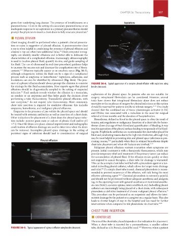
centesis. 8-10 Effusions typically appear as an anechoic space (Fig. 56-5),
although echogenicity within the fluid can be a sign of a complicated
process such as empyema or hemothorax. Septations, adhesions, and
3
loculations can also be identified by ultrasound (Fig. 56-6). The pres-
ence of a pleural effusion should always prompt the clinician to consider FIGURE 56-6. Typical appearance of a complex pleural effusion with septations using
the etiology for the fluid accumulation. Most experts agree that pleural beside ultrasound.
effusions should be diagnostically sampled in the setting of suspected
infection. Fluid analysis reveals whether the effusion is a transudate,
33
an exudate or an empyema and thus helps guide the decision about exploration of the pleural space. In patients who are not suitable for
surgery, intrapleural fibrinolysis can be considered. However, several
performing a tube thoracostomy. Transudative pleural effusions, with
rare exception, do not require tube thoracostomy. More commonly, trials have shown that intrapleural fibrinolysis alone does not reduce
11
mortality or the incidence of surgery for pleural infections so this option
chest tube insertion is required for exudative effusions: this includes 12-14
empyema, hemothorax, and malignant pleural effusions. should be reserved for patients unable to tolerate surgery. One study
showed that the combined use of tissue plasminogen activator (t-PA)
Empyema is the presence of pus within the pleural space and should
be treated with systemic antibiotics as well as insertion of a chest drain. and DNAse was associated with a reduction in the need for surgical
referral at three months and the duration of hospitalization.
Other indications for placement of a chest drain for pleural space infec-
Hemothorax, defined as blood in the pleural space, is often the result of
tion include: positive gram stain or culture of pleural fluid and/or pH trauma, anticoagulation or malignancy. Insertion of a chest tube for hemo-
<7.2. Once the drain is in place, clinical improvement and radiographic
confirmation of effusion drainage are used to determine when the drain thorax allows drainage of fresh blood and quantification of bleeding; it may
result in apposition of the pleural surfaces leading to tamponade of the bleed-
can be removed. Incomplete pleural space drainage in the setting of
persistent signs of infection should lead to consideration of surgical ing site. Prophylactic antibiotics are recommended for chest tubes placed for
blunt and penetrating trauma due to the high risk of infection, although they
may be most helpful in preventing skin and pleural space infections in pen-
etrating trauma, 15,16 particularly when there is retained hemothorax despite
chest tube placement and when rib fractures are involved. 17-19
Malignant pleural effusions warrant evacuation when symptoms are
present. Initial treatment is with a therapeutic thoracentesis, which may
provide temporary relief until treatment of the primary tumor can reduce
the accumulation of pleural fluid. If the effusion recurs quickly or does
not respond to cancer therapies, a chest tube for drainage is warranted.
This can be a simple chest tube instilled with a pleuredesis agent or a tun-
neled indwelling pleural catheter, which allows for longer-term drainage
and relief. If a simple chest tube is used, chemical pleurodesis is recom-
20
mended to prevent recurrence of the effusion, with talc being the most
effective sclerosing agent. 20,21 Chemical pleurodesis is extremely painful,
and should not be performed without adequate anesthesia and analgesia
(eg, in the operating room with general anesthesia or monitored anesthe-
sia care (MAC), systemic opiates, intercostal block, etc). Indwelling pleural
catheters are increasingly being placed of a chest drain, with subsequent
pleurodesis as definitive treatment of chronic malignant pleural effusions.
36
This procedure can be performed as an outpatient and allows for inter-
mittent drainage by the patient or a caregiver on a regular basis. This also
leads to shorter length of stay in the hospital and less need for further
interventions when compared to talc pleurodesis via chest tube. 22,23
CHEST TUBE INSERTION
■ CATHETER SIZE
The size of the chest tube placed depends on the indication for placement.
When a chest tube is inserted for a pneumothorax, a smaller caliber
2,29
FIGURE 56-5. Typical appearance of a pleural effusion using beside ultrasound. tube, defined as ≤14 French, should be used. However, when a patient
section04.indd 507 1/23/2015 2:20:21 PM