
Page 867 - Hall et al (2015) Principles of Critical Care-McGraw-Hill
P. 867
598 PART 5: Infectious Disorders
throat, half develop a tender or swollen neck, and 80% will develop Resolution of the infection always requires device removal when there
pleuropulmonary disease. Patients with pylephlebitis typically have is deep pocket involvement or where S aureus is the cause. Intravenous
48
abdominal pain and abnormal liver function tests. Those with septic antibiotics alone may be effective in all other cases of uncomplicated
pelvic thrombophlebitis usually have persistent fever and 50% present infection. Treatment regimens generally are the same used to treat IE. A
with pelvic pain. 49 2-week course of treatment may be adequate in patients who do not have
■ ETIOLOGY S aureus infection, evidence of IE, and have had all hardware removed. A
57
new pacemaker may be placed once the patient’s bacteremia has cleared.
of cases. In fact, the isolation of fusobacterium from blood cultures ■ ARTERIAL GRAFT INFECTIONS
Lemierre syndrome is caused by fusobacterium in more than 80%
should always raise the question of postanginal sepsis. Secondary The literature reports the infection rate for arterial grafts to be between
infection may occur due to S aureus or other oropharyngeal organisms. 2% and 6%, 58,59 and a reported mortality rate as high as 50%. However,
50
Pylephlebitis is usually due to enteric pathogens such as E coli, Proteus, these reported numbers do not reflect the data regarding abdominal
and Klebsiella, as well as the anaerobes Bacteroides and Clostridia. Septic and thoracic endografts where the infection rate is 0.26% and 4.77%,
pelvic thrombophlebitis has been attributed to enteric pathogens as well respectively. Bruin et al reported a 6-year experience showing the over-
60
as streptococci and staphylococci. 51 all complication rate to be higher with endografts but similar survival
■ DIAGNOSIS AND TREATMENT and infection rates between open and endovascular procedures. Graft
61
infections present on average 8 months after implantation, but have been
Blood cultures are positive in 80% of patients with pylephlebitis but reported to occur as late as 7 to 10 years after graft placement.
usually are not positive in patients with septic pelvic thrombophlebitis As with other intravascular device infections, the etiology and presen-
or postanginal sepsis. Diagnosis is supported by imaging studies with CT tation vary depending on the onset in relation to surgery. Infections that
or MRI having reported sensitivities and specificities of 90% to 100%. 52 occur within 4 months of surgery are considered to be early onset and
Treatment with intravenous antibiotics is usually continued until most often are due to S aureus, whereas late infection is more often due
clinical resolution, which is often 3 to 6 weeks in patients with Lemierre to S epidermidis. Other organisms are encountered less often with only
syndrome or pylephlebitis but usually only 48 to 72 hours after deferves- 14% of infections being polymicrobial. 62
cence in patients with septic pelvic thrombophlebitis. Anticoagulation Patients with early onset infection may present with signs of systemic
may be beneficial in all, but most clearly so in patients with septic pelvic illness, whereas patients with late onset infection often present mostly
thrombophlebitis. Surgical intervention is at times necessary to treat the with signs of graft malfunctioning or poor surgical site wound healing.
primary infection or complications such as abscess formation. Blood cultures are often negative, especially in late onset infection.
CT scan may reveal fluid around the graft, but this may be a normal
early postoperative finding. Technetium or indium scans may be useful
DEVICE-RELATED INFECTIONS as can sinography.
■ PACEMAKER AND DEFIBRILLATOR INFECTIONS venous antibiotics directed against the isolated pathogen.
Definitive treatment involves surgical graft removal along with intra-
as the number of devices implanted has increased 10-fold in recent years ■ CENTRAL VENOUS LINE INFECTIONS
The number of cardiac device–related infections (CDRI) has increased
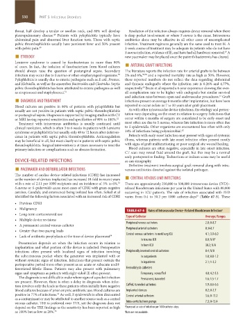
with a rate of 2.11 per 1000 recipients and an incidence of 5% to 6%. There are approximately 250,000 to 500,000 intravenous device (IVD)–
53
S aureus or S epidermidis cause most cases of CDRI with gram-negative related bloodstream infections per year in the United States with 80,000
aerobes, Candida, and enterococci being isolated less often. Sohail et al occurring in ICU patients. The rate of infection associated with IVD
identified the following factors associated with an increased risk of CDRI varies from 0.4 to 30.2 per 1000 catheter-days (Table 67-5). These
63
• Previous CDRI
• Malignancy TABLE 67-5 Rates of Intravascular Device–Related Bloodstream Infection a
• Long-term corticosteroid use Type of Catheter Average; Ranges
• Multiple device revisions
• A permanent central venous catheter Peripheral venous catheters 2.0; 0-8.7
Peripheral arterial catheters 0; 0-8.7
• Greater than two pacing leads
• Lack of antibiotic prophylaxis at the time of device placement 54 Central venous catheters in med/surg ICU 4.1; 3.9-6.0
In trauma ICU 8.0; N/A b
Presentation depends on when the infection occurs in relation to
implantation and what portion of the device is infected. Perioperative In burn ICU 30.2; N/A
infections often present with localized signs of infection involving Peripherally inserted central catheters 0.4; N/A
the subcutaneous pocket where the generator was implanted with or In outpatients 1.0; 0.8-1.2
without systemic signs of infection. Infections that present outside the In inpatients 2.1; 1-3.2
perioperative period more often present as an acute or subacute undif-
ferentiated febrile illness. Patients may also present with pulmonary Hemodialysis catheters
signs and symptoms as patients with right-sided IE often present. Temporary, noncuffed 4.8; 4.2-5.3
The diagnosis is not difficult to make where signs of a pocket infection Permanent, tunneled 1.6; 1.5-1.7
are present. However, there is often a delay in diagnosis when infec-
tion inv olves only the leads as these patients often initially have negative Cuffed, tunneled catheters 1.9; 0.6-6.6
blood cultures because of prior empiric antibiotic use. Blood cultures are Implanted devices 0.2; 0-2.7
positive in 77% of infections. As well, S epidermidis is often discounted Central arterial catheters 3.6; 0-13.2
55
as a contaminant or may be attributed to another source such as a central
venous catheter. TEE is preferred over TTE, yet the diagnosis does not Intra-aortic balloon pumps 7.3; 0-15.4
depend on the TEE findings as the sensitivity has been reported as high a Expressed as rate of infection per 1000 catheter-days.
as 100% but as low as 20%. 56 b Data are not available.
section05_c61-73.indd 598 1/23/2015 12:48:01 PM