
Page 900 - Hall et al (2015) Principles of Critical Care-McGraw-Hill
P. 900
CHAPTER 69: Human Immunodeficiency Virus (HIV) and AIDS in the Intensive Care Unit 631
https://kat.cr/user/tahir99/
FIGURE 69-6. Characteristic foamy honeycomb material seen in alveolar spaces in
Pneumocystis pneumonia (H&E stain, × 3100).
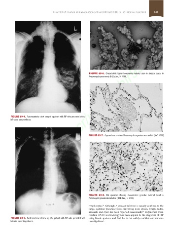
FIGURE 69-4. Posteroanterior chest x-ray of a patient with PJP who presented with a
left-sided pneumothorax.
FIGURE 69-7. Cup-and-saucer-shaped Pneumocystis organisms seen on BAL (GMS 3 100).
FIGURE 69-8. BAL specimen showing characteristic granular material found in
Pneumocystis pneumonia infection (H&E stain, × 3100).
lymphocytes. Although P jirovecii infection is usually confined to the
80
lungs, systemic pneumocystosis (involving liver, spleen, lymph nodes,
adrenals, and eyes) has been reported occasionally. Polymerase chain
81
reaction (PCR) methodology has been applied to the diagnosis of PJP
FIGURE 69-5. Posteroanterior chest x-ray of a patient with PJP who presented with using blood, sputum, and BAL but is not widely available and remains
bilateral upper lung disease. investigational.
section05_c61-73.indd 631 1/23/2015 12:48:19 PM