
Page 954 - Hall et al (2015) Principles of Critical Care-McGraw-Hill
P. 954
CHAPTER 73: Life-Threatening Infections of the Head, Neck, and Upper Respiratory Tract 685
hoarseness, and respiratory distress. Respirations are noisy, often
accompanied by chest wall retractions and inspiratory and expiratory
wheezing. Nasal discharge and pharyngeal injection are common, but
the epiglottis and supraglottic structures appear normal. Fever and mal-
aise are present as part of the upper respiratory viral syndrome. A lateral
radiograph of the neck can be helpful by showing the characteristic
infraglottic narrowing. Management is similar to that for supraglottic
laryngitis, including humidification, hydration, oxygen administration,
and antibiotic therapy for secondary bacterial infection. Use of sedatives
and narcotics, which suppress the cough reflex, is to be avoided.
Inhalational or oral steroids are of proven benefit. Occasionally, an
22
artificial airway is required for 2 to 5 days or more. Extubation is some-
times difficult because of additional edema secondary to the endotra-
cheal tube itself. It seems reasonable that if the patient fails extubation, a
tracheostomy should be considered instead of reintubation.
■ PERICRANIAL INFECTIONS
Contiguous Extension From Sinusitis and Mastoiditis: Fortunately, sup-
purative and life-threatening complications of acute and chronic
sinusitis or mastoiditis have become relatively infrequent in the post-
antibiotic era. However, because of the unique pericranial location of
these air spaces and the rich vascular supply in this region, contiguous
spread of infection may extend intracranially via the diploic veins and
result in serious complications such as meningitis, brain abscess, sub-
dural or epidural empyema, osteomyelitis of the skull, and cavernous
and other cortical venous sinus thrombosis (Fig. 73-2). The clinical
23
spectrum of such complications may be quite varied (Table 73-3).
Since the roof of the frontal and ethmoidal sinuses forms the anterior
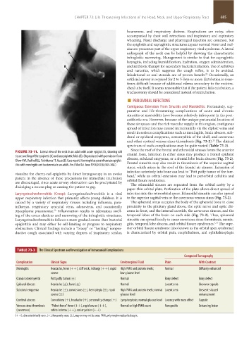
FIGURE 73-11. Lateral view of the neck in an adult with acute epiglottitis, showing soft cranial fossa, infection in either sinus may produce a frontal epidural
tissue swelling of the epiglottis (A) and aryepiglottic folds (B). (Reproduced with permission from abscess, subdural empyema, or a frontal lobe brain abscess (Fig. 73-2).
Chow AW, Bushkell LL, Yoshikawa TT, Guze LB. Case report. Haemophilus parainfluenzae epiglot- Frontal sinusitis may also result in thrombosis of the superior sagittal
titis with meningitis and bacteremia in an adult. Am J Med Sci. June 1974;267(6):365-368.)
sinus, which arises in the roof of the frontal air sinuses. Extension of
infection anteriorly into bone can lead to “Pott puffy tumor of the fore-
visualize the cherry-red epiglottis by direct laryngoscopy in an awake head,” while an orbital extension may lead to periorbital cellulitis and
patient in the absence of these precautions for immediate intubation orbital fissure syndromes.
are discouraged, since acute airway obstruction can be precipitated by The ethmoidal sinuses are separated from the orbital cavity by a
dislodging a mucus plug or causing the patient to gag.
paper-thin orbital plate. Perforation of the plate allows direct spread of
Laryngotracheobronchitis (Croup): Laryngotracheobronchitis is a viral infection into the retroorbital space. Ethmoidal sinusitis can also spread
upper respiratory infection that primarily affects young children. It is to the superior sagittal vein or the cavernous venous sinus (Fig. 73-2).
caused by a variety of respiratory viruses including influenza, para- The sphenoid sinus occupies the body of the sphenoid bone in close
influenza, respiratory syncytial virus, adenovirus, and occasionally proximity to the pituitary gland above, the optic nerve and optic chi-
Mycoplasma pneumoniae. Inflammation results in edematous swell- asma in front, and the internal carotids, the cavernous sinuses, and the
20
ing of the conus elasticus and narrowing of the infraglottic structures. temporal lobes of the brain on each side (Fig. 73-2). Thus, sphenoid
Laryngotracheobronchitis follows a more gradual course than bacterial sinusitis can spread locally to cause cavernous sinus thrombosis, menin-
2,24
epiglottitis and may either be self-limiting or progress to respiratory gitis, temporal lobe abscess, and orbital fissure syndromes. The supe-
obstruction. Clinical findings include a “brassy” or “barking” nonpro- rior orbital fissure syndrome (also known as the orbital apex syndrome)
ductive cough associated with varying degrees of inspiratory stridor, is characterized by orbital pain, exophthalmos, and ophthalmoplegia
TABLE 73-3 The Clinical Spectrum and Investigation of Intracranial Complications
Computed Tomography
Complication Clinical Signs Cerebrospinal Fluid Plain With Contrast
Meningitis Headache, fever (++), stiff neck, lethargy (++), rapid High PMN and protein levels; Normal Diffusely enhanced
course low glucose level
Cranial osteomyelitis Pott puffy tumor (±) Normal Bony defect Bony defect
Epidural abscess Headache (±), fever (±) Normal Lucent area Biconvex capsule
Subdural empyema Headache (±), convulsions (±), hemiplegia (±), rapid High PMN and protein levels; normal Lucent area Crescent-shaped
course (±) glucose level enhancement
Cerebral abscess Convulsions (+), headache (+), personality change (+) Lymphocytosis; normal glucose level Lucency with mass effect Capsule
Venous sinus thrombosis “Picket-fence” fever (++), rapid course (++), Normal or high PMN count Nonspecific Enhancing lesion
(cavernous) orbital edema (++), ocular palsies (++)
(++), characteristically seen; (+), frequently seen; (±), may or may not be seen; PMN, polymorphonuclear leukocyte.
section05_c61-73.indd 685 1/23/2015 12:49:12 PM