
Page 956 - Hall et al (2015) Principles of Critical Care-McGraw-Hill
P. 956
CHAPTER 73: Life-Threatening Infections of the Head, Neck, and Upper Respiratory Tract 687
syringe for aspiration of loculated pus through an extraoral approach.
After the skin is cleansed, pus is aspirated into the syringe. All air is
carefully expressed, and the needle tip is inserted into a rubber stopper.
This allows the exclusion of air, and the specimen can then be trans-
ported directly to the laboratory. This method of specimen collection
is superior to using swabs. If a swab is used, it should be saturated with
purulent material and inserted into a commercially available transport
tube specifically designed to transport swabs under anaerobic condi-
tions. An additional swab should be taken for Gram staining. The Gram
stain is particularly useful in the assessment of head and neck infec-
tions because a polymicrobial flora is generally present, and anaerobic
bacteria may require 48 hours or longer for growth. The microscopic
morphology of some of the bacteria may be characteristic enough to
suggest a provisional diagnosis and, ultimately, therapy. Infected tissues
obtained intraoperatively are also suitable for anaerobic and aerobic
processing, provided that care is taken to prevent contamination by the
normal resident flora.
Apart from routine culture and special stains for examination of direct
smears, specimens may also be collected for histopathologic examina-
tion and direct detection of microbial antigens using immunological
or molecular techniques. Nucleic acid amplification methods such
34
as polymerase chain reaction (PCR) and sequence-based analysis are
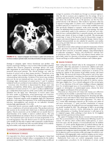
FIGURE 73-12. Computed tomographic scan of the head in a patient with cavernous sinus particularly suited for detection of fastidious microorganisms, certain
thrombosis secondary to sphenoid sinusitis. Arrow indicates thrombus in the right cavernous sinus. viruses and fungi, as well as antibiotic resistance and virulence genes.
■ IMAGING TECHNIQUES
drainage is adequate, septic venous thrombosis may produce only
transient neurologic findings or may be silent. If the thrombus outstrips Plain radiographs have limited value in the management of critically
collateral flow, however, progressive neurologic deficits will result, ill patients, other than placement of intravenous catheters and endo-
with impairment of consciousness, focal or generalized seizures, and tracheal or nasogastric intubation. An exception is a lateral radiograph
increased intracranial pressure. The clinical findings vary with the of the neck, which may demonstrate compression or deviation of the
location of cortical veins or dural sinuses involved. Thrombosis of the tracheal air column or the presence of gas within necrotic soft tissues
superior sagittal sinus produces bilateral leg weakness and may cause (Fig. 73-10). The normal soft tissues of the posterior wall of the hypo-
communicating hydrocephalus. Occlusion of the lateral sinus produces pharynx are approximately 5 mm deep, less than one-third the diameter
pain over the ear and mastoid and may cause edema over the mastoid of the fourth cervical vertebra (C4). The retropharyngeal soft tissues
(Griesinger sign). Involvement of cranial nerves V and VI produces should be approximately two-thirds the width of C4, and the retrotra-
ipsilateral facial pain and lateral rectus weakness (Gradenigo syndrome). cheal space slightly less. Thus, a lateral radiograph of the cervical spine
Cavernous sinus thrombosis is characterized by abrupt onset with or a CT can determine if the soft tissue swelling or abscess originated
diplopia, photophobia, orbital edema, and progressive exophthalmos. from the retropharyngeal space or the prevertebral space. The former
Involvement of cranial nerves III, IV, V, and VI produces ophthalmople- suggests an odontogenic or oropharyngeal source, whereas the latter
gia, a midposition fixed pupil, loss of the corneal reflex, and diminished likely suggests involvement of the cervical spine.
sensation over the upper face. Obstruction of venous return from Ultrasound can characterize soft tissue neck masses and collections
the retina results in papilledema, retinal hemorrhage, and visual loss. but is limited by its inability to penetrate bone or air-filled structures.
Contrast-enhanced CT (Fig. 73-12) and MRI are the imaging modalities CT or MRI are the best imaging techniques for detecting and delin-
of choice. Treatment requires early recognition, high-dose intravenous eating the source and extent of deep fascial space infections of the head
7
antibiotics, and surgical decompression of the underlying predisposing and neck and pericranial or intracranial suppuration. The choice of
infection. Anticoagulation and steroids are not indicated. Mortality a CT versus MRI examination depends on the location and nature of
remains high, approximately 15% to 30%. soft tissue involvement. CT gives excellent visualization of osseous
Intracranial mycotic aneurysm usually results from septic emboliza- structures, particularly the temporal bones and paranasal sinuses, which
tion as a complication of bacterial endocarditis. This produces infection are poorly visualized by MRI. The advantage of MRI is in providing
and necrosis in the arterial wall, which leads to dilation and possible soft tissue contrast resolution, further delineating the extent of soft
rupture. Mycotic aneurysms can be multiple and are usually found on tissue inflammation or bleeding. Normal anatomy is well depicted by
distal branches of the middle or anterior cerebral arteries. The early T1-weighted images, while pathology is best shown by T2-weighted
7
clinical manifestations are similar to those of cerebral emboli and infarc- images and after gadolinium enhancement.
tion. The weakened vessel may be seen to increase progressively in size The typical CT finding in brain abscess is an area of decreased attenu-
on serial angiograms. Since the clinical course of a mycotic aneurysm is ation that is surrounded by a ring of enhancement following injection of
quite variable and the risk of rupture with catastrophic cerebral hemor- contrast. CT will also detect cerebral edema, hydrocephalus, an associ-
rhage cannot be predicted even after successful therapy of the underly- ated mass effect, and the presence of extracranial infection. In subdural
ing endocarditis, early surgical intervention is advised. empyema, CT reveals inward displacement of cerebral substance due to
an extracerebral mass. In epidural abscess, CT demonstrates a thick and
circumscribed area of diminished density associated with extracerebral dis-
DIAGNOSTIC CONSIDERATIONS placement and contiguous cranial osteomyelitis. MRI is particularly useful
■ MICROBIOLOGIC TECHNIQUES for the detection and characterization of the early stages of cerebritis or epi-
dural abscess. MRI angiography is also useful for imaging vascular lesions,
It is imperative that clinical specimens for the diagnosis of deep head such as jugular thrombophlebitis and cranial septic venous thrombosis. 23
and neck infections be obtained without contamination by the resident Radionuclide brain scans and cerebral angiography remain useful
34
oronasopharyngeal flora. This is best accomplished using a needle and as complementary procedures for the localization of certain central
section05_c61-73.indd 687 1/23/2015 12:49:13 PM