
Page 973 - Hall et al (2015) Principles of Critical Care-McGraw-Hill
P. 973
704 PART 5: Infectious Disorders
The bacteria that populate the GI tract are varied, depending on the
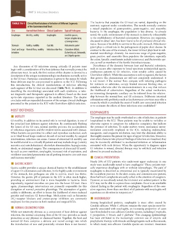
TABLE 76-1 Clinical Manifestations of Infection of Different Segments
of the Gastrointestinal Tract anatomic segment under consideration. The mouth normally contains
a mixed population of gram-positive, gram-negative, and anaerobic
Site Normal Host Defense Clinical Syndrome Typical Pathogens bacteria. In the esophagus, the population is less diverse. As already
Esophagus Motility, acidity Esophagitis Candida species, noted, the acidic environment of the stomach is distinctly inhospitable
herpes simplex virus, to the establishment of bacterial colonization. However, one organism,
cytomegalovirus discussed in detail later, has been found to be of profound clinical rele-
vance. Because of its ability to survive in the acidic stomach, Helicobacter
Stomach Acidity, motility Gastritis Helicobacter pylori
pylori plays a critical role in the pathogenesis of peptic ulcer disease. In
Small and large Normal flora, motility Infectious diarrhea Clostridium difficile, contrast to the case of the stomach, the lower GI tract plays host to sub-
intestine Escherichia coli, Salmonella, stantial microbiologic diversity. An enormous range of gram-negative,
and Shigella species gram-positive, and anaerobic flora populates the intestines, especially
the colon. Specific constituents include enterococci and Bacteroides spe-
cies, as well as members of the family Enterobacteriaceae.
Any discussion of GI infections among critically ill patients must Disturbance of the dynamic between host and bacterial colonizers,
begin with a consideration of the host defenses that normally protect the such as occurs after exposure to broad-spectrum antimicrobial agents,
alimentary tract. As such, the first section of this chapter is devoted to a predisposes patients to GI infection, most notably colitis caused by
description of the unique nonimmunologic mechanisms normally active Clostridium difficile. While this association is well recognized, the factors
in the GI tract. Particular consideration is given to the means by which that govern this phenomenon are still not completely understood. It
these defenses may be compromised in patients in the ICU. Following is not known if the normal flora compete with infecting pathogens
this introduction, the clinical manifestations of infection affecting for nutrients or substrates, occupy limited mucosal binding sites, or
each segment of the GI tract are discussed (Table 76-1). In addition to somehow otherwise alter the microenvironment in a way that reduces
describing the microbiology associated with each syndrome, a ratio- the likelihood of colonization. Regardless of the actual mechanism,
nal diagnostic and therapeutic approach is offered, based on the most an interesting therapeutic corollary can be inferred from the relation-
up-to-date experience reported in the medical literature. The chapter ship between the normal host and GI colonizers. Deliberate intestinal
concludes with an expanded discussion of the unique clinical challenges colonization with probiotics such as Saccharomyces cerevisiae may offer a
presented by the patient in the ICU with Clostridium difficile infection. means by which to preclude the onset of health care–associated infection
or to attenuate the effects of these infections once established. 2
HOST DEFENSES ESOPHAGITIS
■ MOTILITY The esophagus may be easily overlooked as a site of infection in patients
GI motility, in addition to its central role in normal digestion, is one of hospitalized in the ICU. These patients may be unable to verbalize or
the principal host defenses against infection. By continuously flushing otherwise express to caregivers the subjective complaints that indicate
the lumen of the GI tract, normal motility prevents the accumulation the presence of infection. To make matters worse, mechanical instru-
of infectious organisms and the virulent toxins associated with disease. mentation commonly employed in the ICU, including endotracheal,
When bacteria are permitted to collect and reproduce unchecked, such nasogastric, and orogastric intubation, may limit the clinician’s ability to
as in blind bowel loops rendered devoid of normal motility by surgical thoroughly examine the patient for signs of upper GI infection. Moreover,
interventions, infection can ensue. Causes of abnormal GI motility can even when characteristic physical findings of infection are visualized, they
be multifactorial in a critically ill patient and may include drugs (notably may be incorrectly attributed to mechanical irritation or inflammation
narcotics and catecholamines), electrolyte abnormalities, hypoglycemia, associated with such devices. When the opportunity to diagnose upper
shock, or abdominal surgery. The consequences of abnormal GI motil- GI infection is missed, directed therapy may be withheld and infection
ity such as poor nutrition, esophagitis, increased risk of aspiration, and allowed to proceed unchecked.
ventilator-associated pneumonia can all prolong intensive care unit stays
and increase mortality. 1 ■ CLINICAL PRESENTATION
■ GASTRIC ACIDITY Nearly 20% of ICU patients who underwent upper endoscopy in one
3
study were incidentally noted to have esophagitis. These patients typi-
Gastric acidity provides a unique chemical barrier to the establishment cally experience dysphagia with or without odynophagia. The pain of
of upper GI colonization and infection. In the highly acidic environment esophagitis is described as retrosternal and is typically exacerbated by
of the stomach, few pathogens are able to survive, much less thrive. the recumbent position. In the alert, awake, and communicative patient,
However, the gastric pH of patients in the ICU is often much higher, these hallmark complaints are easily called to the attention of caregivers.
providing an environment that is more hospitable to bacteria. More However, as was already noted, the intubated and sedated patient in the
importantly, ingested microbes can pass into the lower GI tract. Once ICU may not be able to express these complaints. Fever is an unreliable
again, pharmacologic interventions are primarily responsible for this clinical finding in the patient with esophagitis. Regardless of the caus-
disruption of normal protective physiology. The attenuation of gastric ative organism, fewer than one-third of all patients with esophagitis will
acidity is deliberate, an effort to lessen the likelihood of stress-induced experience an elevation in temperature. 4
gastritis and resultant GI hemorrhage. Medications such as histamine ■
(H )-receptor blockers and proton-pump inhibitors are commonly MICROBIOLOGY
2
employed for this practice in both medical and surgical ICUs. Among hospitalized patients, esophagitis is most often caused by
■ NORMAL COLONIZING FLORA Candida albicans. While C albicans remains the yeast species most fre-
quently associated with esophagitis, an increasing proportion of cases
While not intuitively obvious as a component of host defense against have been linked to non-albicans Candida species, including C tropicalis,
5
infection, the normal colonizing flora of the GI tract provides as much C parapsilosis, C krusei, and C glabrata. This changing epidemiology
protection as any physical or chemical barrier. Together, the host and has been attributed to the increasingly common use of empiric and
normal GI flora comprise a delicate and varied ecology into which prophylactic therapy with triazole antifungal agents such as fluconazole,
the introduction of new and potentially virulent flora is not favored. to which many non-albicans Candida species are resistant. Awareness
section05_c74-81.indd 704 1/23/2015 12:37:28 PM