
Page 121 - Clinical Anatomy
P. 121
ECA2 7/18/06 6:42 PM Page 106
106 The abdomen and pelvis
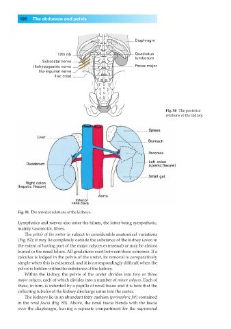
Diaphragm
12th rib Quadratus
lumborum
Subcostal nerve
Iliohypogastric nerve Psoas major
Ilio-inguinal nerve
Iliac crest
Fig. 80◊The posterior
relations of the kidney.
Fig. 81◊The anterior relations of the kidneys.
Lymphatics and nerves also enter the hilum, the latter being sympathetic,
mainly vasomotor, fibres.
The pelvis of the ureter is subject to considerable anatomical variations
(Fig. 82); it may lie completely outside the substance of the kidney (even to
the extent of having part of the major calyces extrarenal) or may be almost
buried in the renal hilum. All gradations exist between these extremes. If a
calculus is lodged in the pelvis of the ureter, its removal is comparatively
simple when this is extrarenal, and it is correspondingly difficult when the
pelvis is hidden within the substance of the kidney.
Within the kidney, the pelvis of the ureter divides into two or three
major calyces, each of which divides into a number of minor calyces. Each of
these, in turn, is indented by a papilla of renal tissue and it is here that the
collecting tubules of the kidney discharge urine into the ureter.
The kidneys lie in an abundant fatty cushion (perinephric fat) contained
in the renal fascia (Fig. 83). Above, the renal fascia blends with the fascia
over the diaphragm, leaving a separate compartment for the suprarenal